
Research
Surgical simulators provide a safe environment where novice surgeons can acquire their surgical skills. Depending on the technical implementation, one can distinguish between different simulator types.
Model based or “Low Fidelity” simulators (also known as “Box Trainers”) allow the usage of original surgical instruments on simple physical models or specimens. This affordable simulator type has limited feedback possibilities resulting in the need of immediate (subjective) trainee assessment.
Computer based or “Virtual Reality” (VR) simulators provide a detailed visualization of morphological data as well as more feedback possibilities than box trainers. The haptic feedback is usually insufficient due to complex algorithms used for tissue modelling. In most cases, real surgical instruments are not used and the costs are substantially higher.
Hybrid or “Mixed Reality” (MR) simulators as well as VR trainers belong to the so called “High Fidelity” simulators. The difference between them is that MR trainers are a combination of the aforementioned simulator types. They provide a detailed visualization, enable the usage of original surgical instruments and offer realistic haptic feedback. Only the costs have to be mentioned as a possible limiting factor.
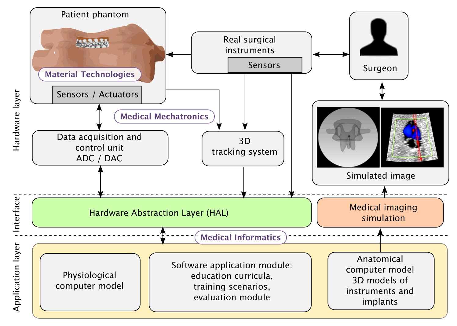
A Hybrid Surgical Simulator combines the advantages of Model-Based and Virtual Reality Simulators and consists of four components:
Patient phantom
Artificial anatomical structures with realistic haptic feedback that also reproduce relevant physiological processes.
Smart surgical instruments
The real surgical instruments, equipped with additional sensors for position tracking and data acquisition.
Data acquisition and control
A unit for data transfer and for controlling additional actuators, e.g. to simulate bleeding.
Software application
Computer models of anatomy and physiology, imaging simulation, education curricula and training scenarios.
The research areas touched by such simulators are multi-disciplinary (as indicated in Figure 1) and includes Material Technologies for the development of artificial anatomical structures, Medical Mechatronics for improving tracking devices and for the development of smart instruments and finally Medical Informatics for the development of algorithms for improved tracking accuracy and registration as well as computer models allowing to simulate anatomical structures and physiological process.
Research Areas
Artificial Bones and Soft Tissue
Artificial bones and soft tissue that reproduce the haptic feedback of human anatomy, validated against human specimens.
Smart Artificial Tissue and Surgical Instruments
Artificial tissue and instruments that sense position, force and deformation, and tissue that moves by itself.
Simulator Validation
Studies with surgeons that prove our simulators are realistic, suitable for teaching and able to assess skill.