

Dynamic Tactile Synthetic Tissue
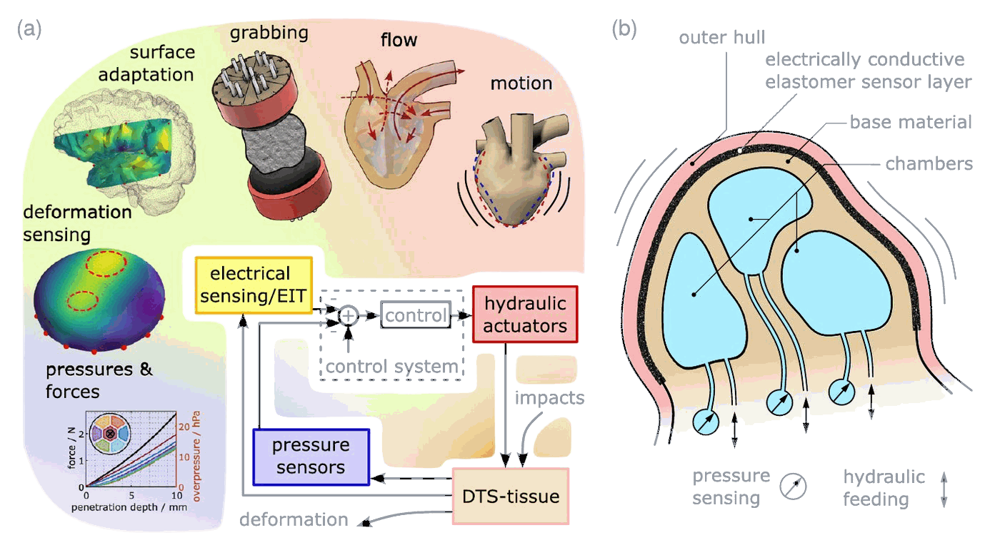
In this project, we developed Dynamic Tactile Synthetic (DTS) Tissue: a fully soft artificial tissue that can both feel and move. It combines hydraulic actuation, pressure sensing and a soft tactile sensor layer in one unit, and is a core building block for dynamic patient phantoms in future hybrid surgical simulators, as well as for tactile soft robotics in general [1].
Motivation
Surgical simulators improve the proficiency of young surgeons and patient safety, but physical patient phantoms are usually static. Real surgery, however, involves moving structures: pulsating vessels, a beating heart, brain swelling, or brain lobes that have to be retracted to open a surgical corridor. Without integrated actuators, an artificial brain cannot perform such retractions, and without integrated sensors, a phantom cannot tell how gently or roughly it was handled. Existing organ simulators with motion capabilities lack exactly this quantitative feedback, which is essential for objective performance evaluation and debriefing.
Tissue that feels and moves
The DTS tissue consists of liquid-filled chambers embedded in a soft silicone base, covered by a thin, electrically conductive sensor layer made of carbon black silicone composite (see Figure 1). This combination provides three functions in one soft unit:
Actuation
Each chamber is filled and emptied hydraulically, so the tissue can inflate, shrink, enclose objects, change its perceived softness and move.
Pressures and forces
Pressure sensors in every chamber measure external impacts and indicate roughly where they occur.
Tactile sensing
Electrical impedance tomography (EIT) of the sensor layer reconstructs where and how strongly the surface is deformed.
All data flow into a control system, which closes the loop between sensing and actuation: the tissue can react to its environment in real time. Since only soft, elastic materials are located in the active region, the tissue feels natural, and its basic structure can be shaped and scaled to almost any geometry. The measurements use our open-source sensor interface for soft detectors in surgical simulators.
Soft tissue gripper
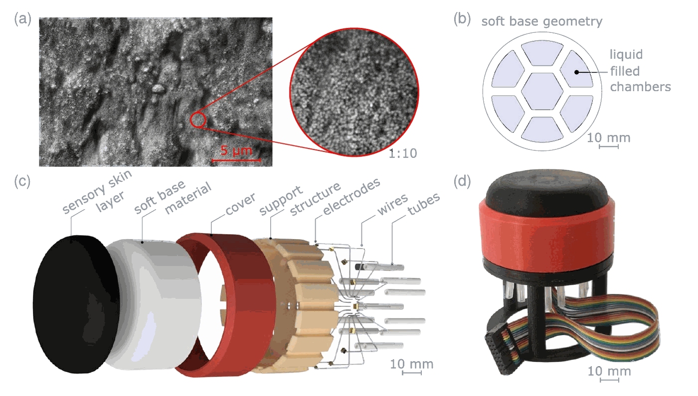
To evaluate the DTS tissue quantitatively, we first built a geometrically well-defined soft gripper with seven chambers (see Figure 2). It was manufactured by silicone casting in 3D-printed molds with water-soluble cores and tested in two softness variants (Ecoflex 10 and 35).
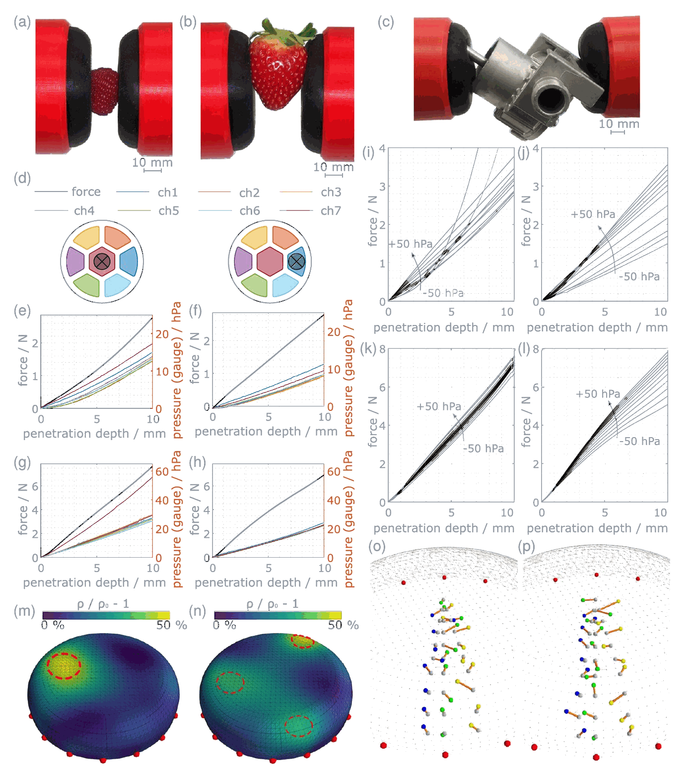
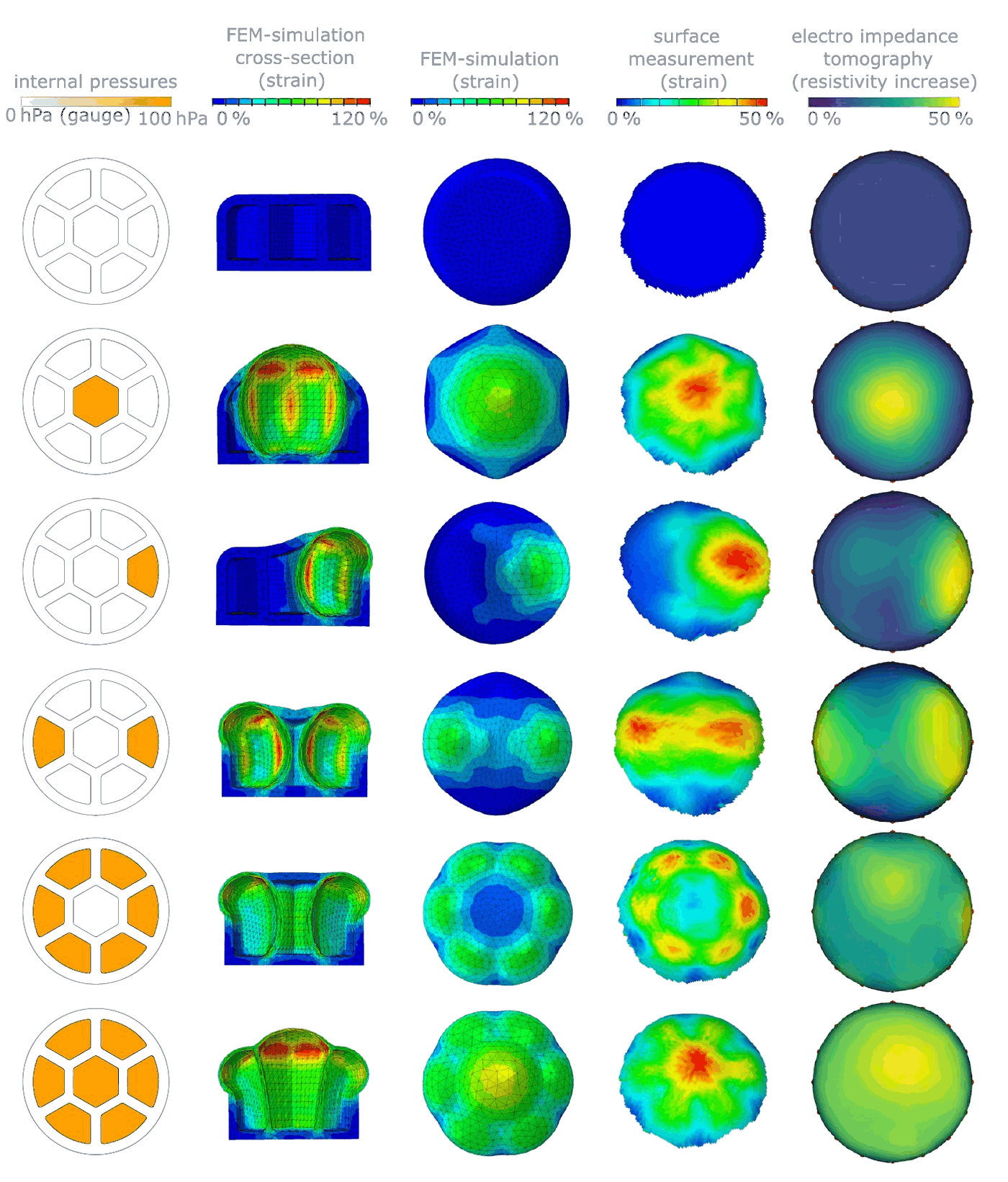
The gripper held a raspberry without damaging it, a strawberry, and a heavy, irregularly shaped metal part of 0.7 kg, adapting its surface to the object (see Figure 3). Forces as small as 0.02 to 0.04 N were detected through the chamber pressures, the softness of the surface could be adjusted via the chamber pressure, and EIT located single touch points with a mean error of about 2.1 to 2.4 mm. Active deformations produced by the chambers were reconstructed by EIT in close agreement with finite element simulations and optical surface measurements (see Figure 4).
Smart artificial brain tissue
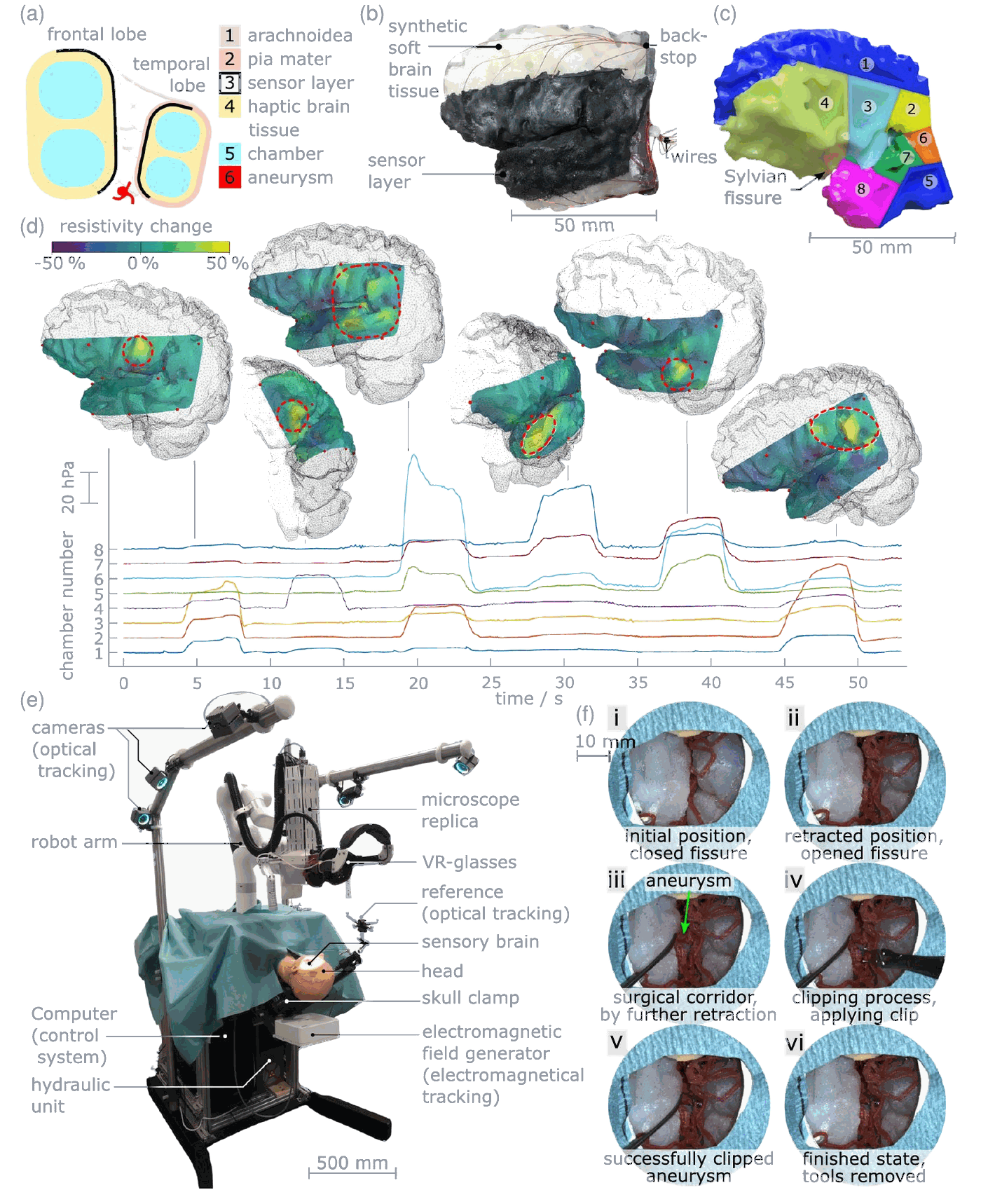
In aneurysm clipping, the surgeon has to open the Sylvian fissure between the frontal and temporal lobe to reach the aneurysm, mobilising the brain lobes without damaging the tissue. We equipped an artificial brain with fluid-filled chambers and a sensor layer following the brain’s folded surface (see Figure 5). Inside a skull model, the chambers move the brain lobes and widen or close the fissure, while the sensor layer shows trainees immediately where and how strongly they stress the brain. Forces from 0.04 N were detectable, and contact points were located with a mean error of about 5.8 mm on the highly curved brain surface.
The sensory brain is part of the hybrid simulator MEDUSA (Medical EDUcation in Surgical Aneurysm Clipping), which combines it with optical instrument tracking, electromagnetic tracking of the soft tissue, and a surgical microscope replica on a robotic arm whose view is shown in VR glasses. Unlike haptic input devices or tracking systems alone, the DTS tissue measures impacts directly in the tissue, without force limits or restrictions on the instruments.
Beating fetal heart
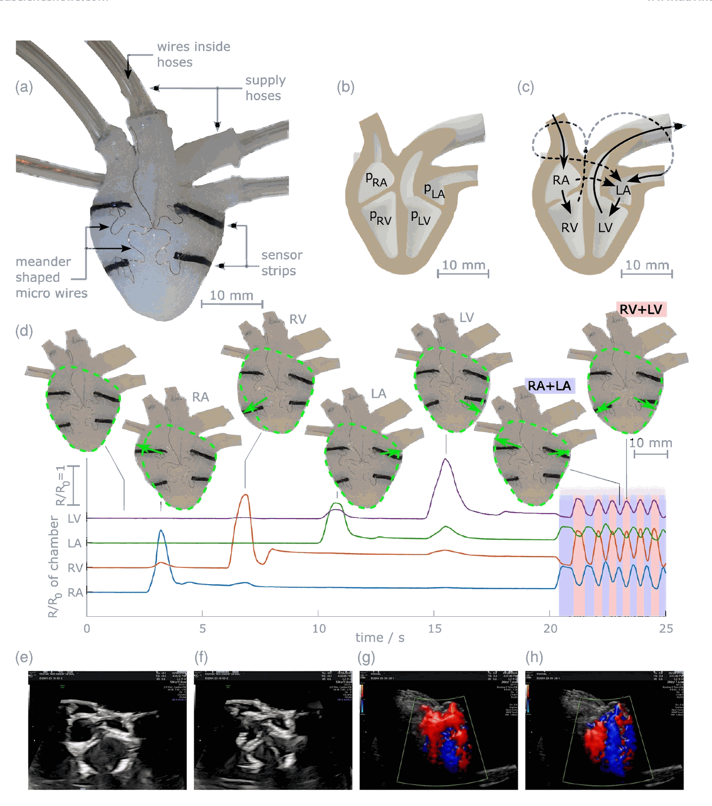
As a further application, we built a beating fetal heart of 22 mm diameter, corresponding to about the 23rd week of pregnancy, with four chambers and one sensor strip each (see Figure 6). The chambers can be driven individually or in a heartbeat pattern, and the sensor strips report the resulting movements. Driven by the closed loop, such a heart could reproduce arbitrary heart rates, arrhythmias or even recorded ECG signals. A variant with connected chambers generates a flow through the heart that is visible in Doppler echocardiography, which makes it useful for ultrasound training and for developing ultrasound imaging algorithms.
Outlook
The DTS tissue brings movement and objective feedback together in haptically realistic artificial anatomy, filling a gap that neither virtual reality, passive phantoms nor biological specimens can close alone. Next steps include a higher sensor resolution, larger movements and miniaturisation for more intricate applications. The tissue builds on our work on carbon black silicone sensors, such as the Smart Artificial Soft Tissue of our laryngeal pacemaker simulator [2].
Partners and funding
This work was carried out in cooperation with the Division of Soft Matter Physics and the Institute of Polymer Product Engineering at Johannes Kepler University Linz and the Soft Materials Lab of the Linz Institute of Technology. It was partly funded by the project MEDUSA within the programme “Innovative Upper Austria 2020” (grant 872604), the project “Fetal Heart Simulator for the Development and Optimization of Ultrasound Imaging Algorithms” (FHSimApUs) within the programme “Digital Health – The Digital Patient Journey” of the Province of Upper Austria (grant 41634460), and the European Research Council Starting Grant GELSYS (grant 757931).
Figures from [1], licensed under CC BY 4.0.
References
- (2024): Carbon black silicone composite in surgical simulation - A detailed electromechanical characterization. In: Sensors and Actuators, A: Physical, vol. 370, pp. 115262, 2024, ISSN: 0924-4247, (Publisher Copyright: © 2024 The Authors).
- (2023): Smart Artificial Soft Tissue - Application to a Hybrid Simulator for Training of Laryngeal Pacemaker Implantation. In: IEEE Transactions on Biomedical Engineering, vol. 70, no. 2, pp. 735–746, 2023, ISSN: 0018-9294, (Publisher Copyright: © 1964-2012 IEEE.).