
Smart Artificial Soft Tissue
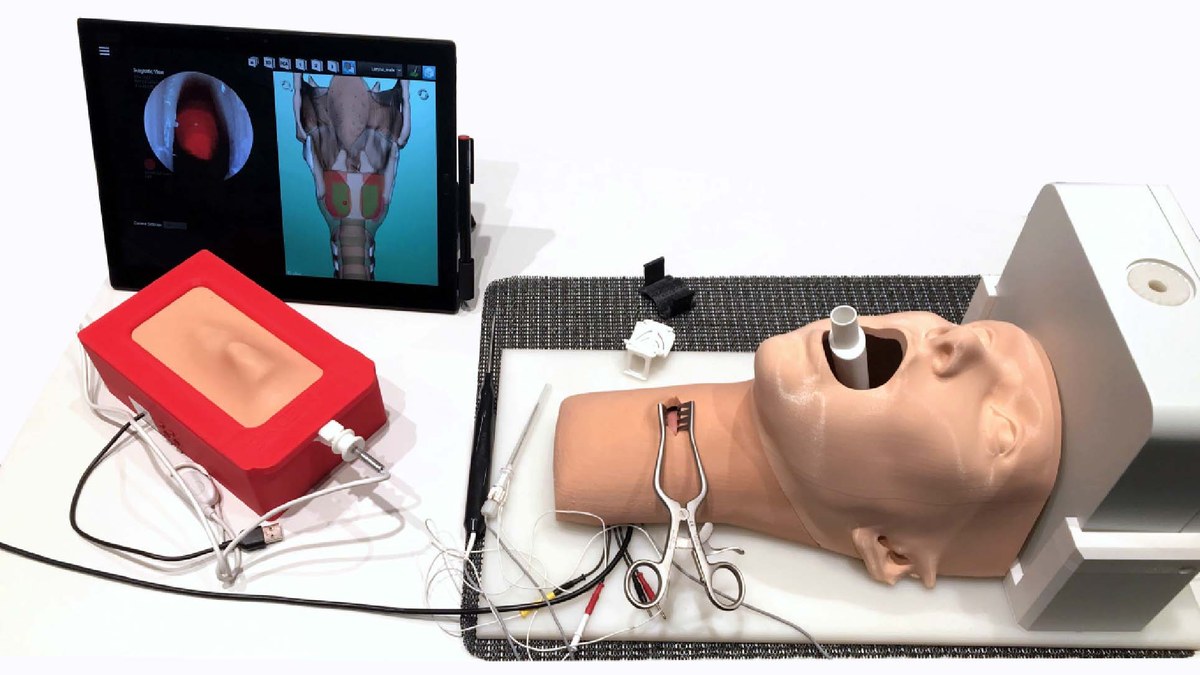
In this project, we developed a hybrid surgical simulator for training the implantation of laryngeal pacemaker electrodes. At its core is a new type of sensing material, the Smart Artificial Soft Tissue: a tissue-mimicking silicone layer that detects the position of conductive surgical instruments inside the phantom, while providing realistic haptic feedback [1], [2].
Motivation
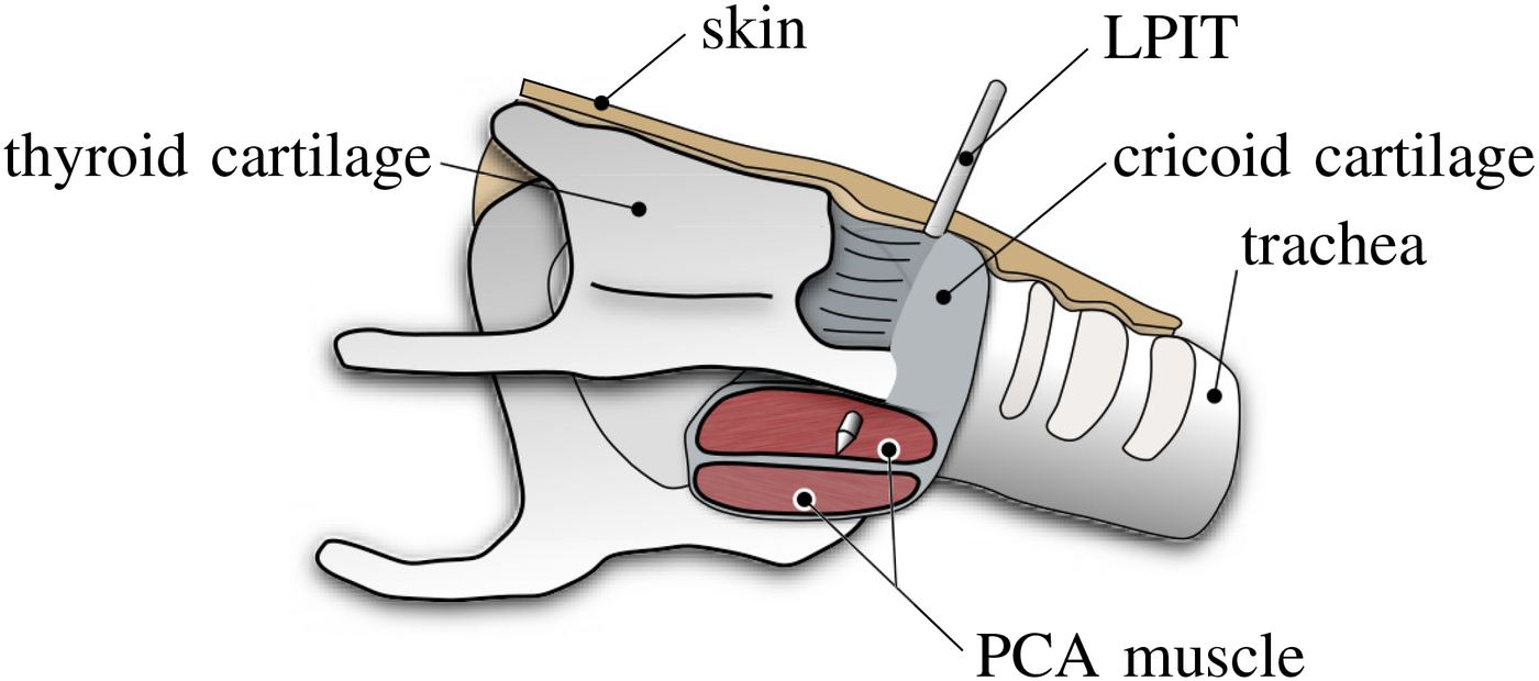
The laryngeal pacemaker (LP) system developed by MED-EL treats patients with bilateral vocal fold paralysis by selectively stimulating the nerve branches that open the vocal folds. During implantation, an LP insertion tool (LPIT) is advanced through the skin, along the cricoid cartilage and through the cricoid lamina into the posterior cricoarytenoid (PCA) muscle (see Figure 1). There, the surgeon has to find a sensitive area, the so-called hotspot, where electrical stimulation opens the vocal fold best. Finding the hotspot and fixing the electrode are the most critical steps of the intervention, and therefore the ones that benefit most from training.
Functional testing and training of such procedures usually rely on animal models, which raises ethical concerns and causes high costs. A simulator is a valuable alternative in the spirit of the 3R principle (replacement, reduction, refinement), but it has to reproduce the haptics of penetrating skin, mucosa, cartilage and muscle, and it has to know where the instrument tip is located inside deformable soft tissue. Conventional optical or electromagnetic tracking systems are poorly suited for this: markers cannot be placed inside the soft tissue, and sensor coils are expensive, alter the haptics and require cabling.
Haptically realistic artificial tissue
Haptic realism was focused on the structures that are actually penetrated by the LPIT: the skin, the cricoid cartilage and the tissue directly surrounding it. The artificial cricoid consists of a 3D-printed TPU shell, thinned to 0.3 mm and coated with a thin layer of super glue on the injection side to reproduce the sharp first force peak, and filled with a urethane rubber mixed with silicone oil to reduce friction. It is embedded in silicone soft tissue and covered by a mesh-reinforced silicone skin (see Figure 2).
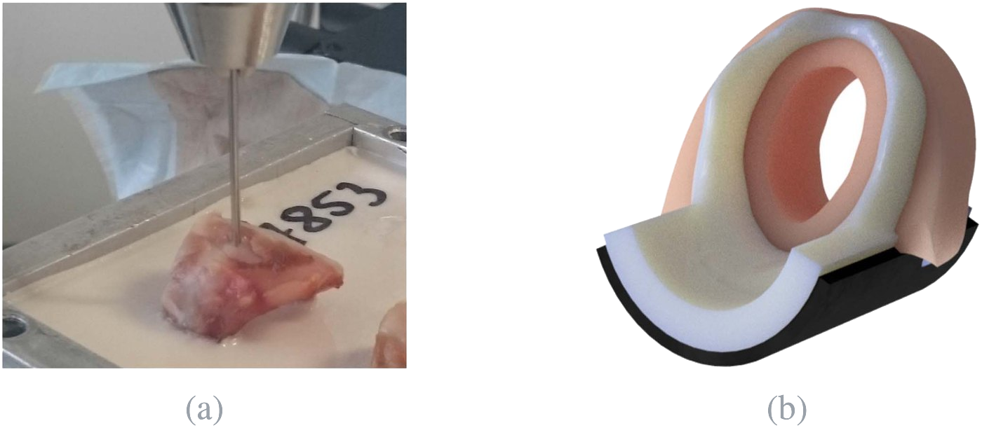
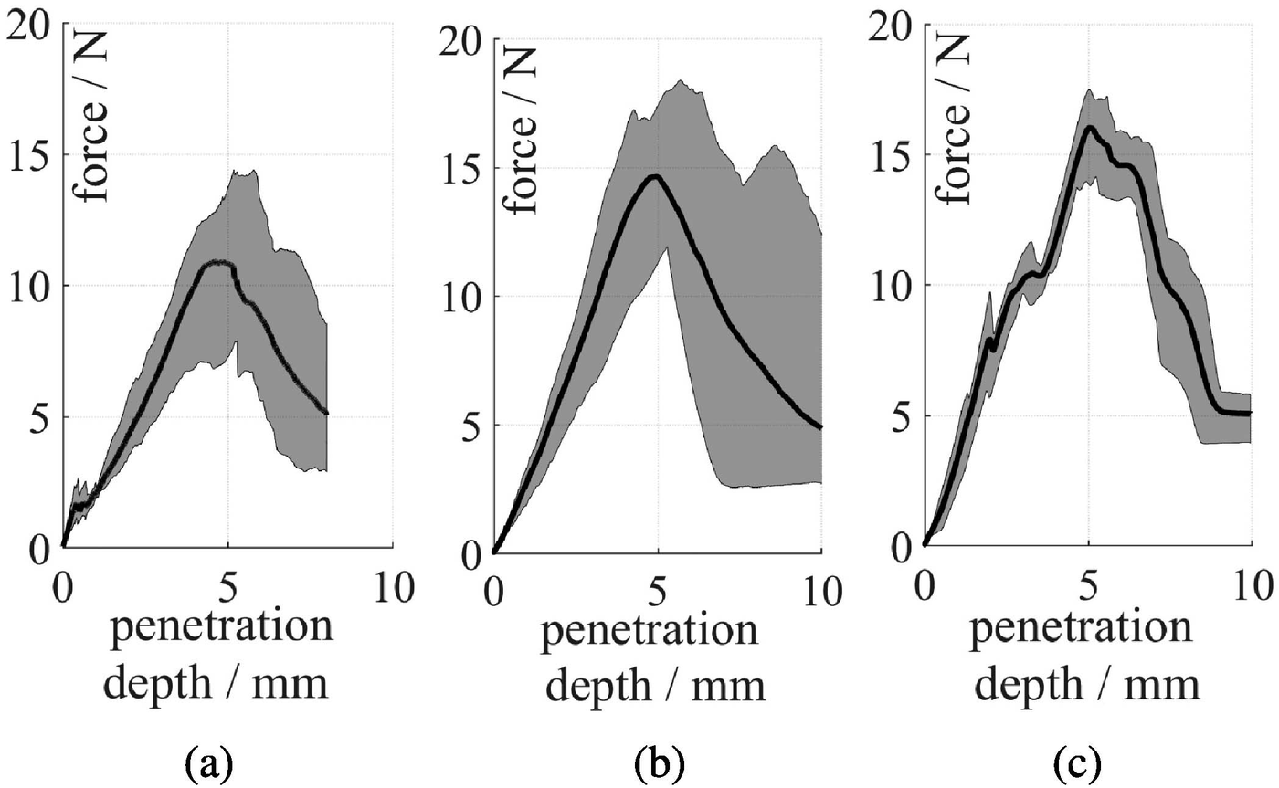
To define the haptic requirements, the forces during penetration of the cricoid lamina were measured on human and porcine larynges with a needle matching the geometry of the LPIT. Human samples showed a maximum mean force of 10.9 N, porcine samples 14.7 N, both with one dominant force peak, and human samples additionally with a small, sharp peak at the beginning. Guided by these measurements and by experienced surgeons, the artificial cricoid reproduces both characteristic peaks and their spacing, with a maximum mean force of 16.0 N (see Figure 3).
Smart Artificial Soft Tissue
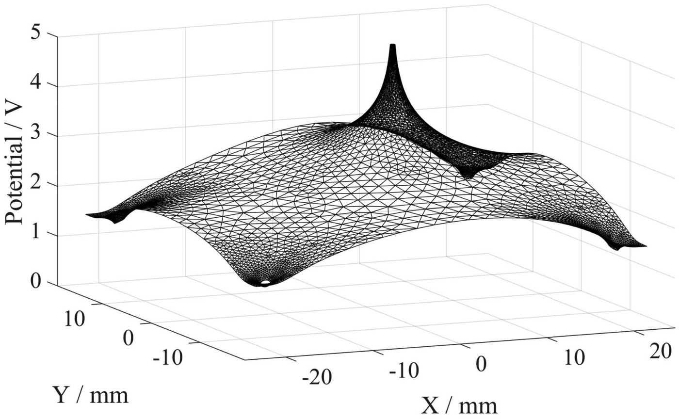
Instead of the PCA muscle, a thin, electrically conductive silicone layer is placed behind the artificial cricoid. Carbon black is mixed into the silicone in a low concentration, so the material stays soft and haptically realistic while becoming conductive. The resulting sensor mat is contacted by four gold-plated electrode pins at its corners. When the voltage-carrying LPIT touches the mat, the current divides among the four pins depending on the tip position, which closely resembles the real situation, where the muscle is stimulated by currents from the LPIT. A finite element model of the electrical potential (see Figure 4) provides the mapping between pin currents and tip position.
The position is reconstructed from ratios of the pin voltages, which makes it independent of the supply voltage and of the varying contact resistance between LPIT tip and mat. A short calibration routine at the start of a session compensates for changes in mat conductivity and pin contact resistance, so each newly inserted sensor mat can be used immediately. Only a low-cost microcontroller is needed for the measurement.
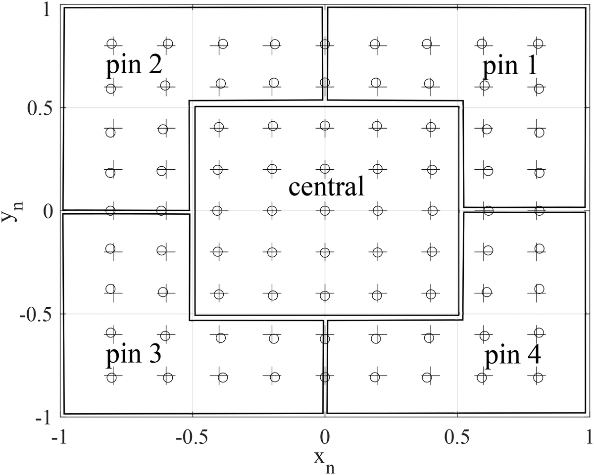
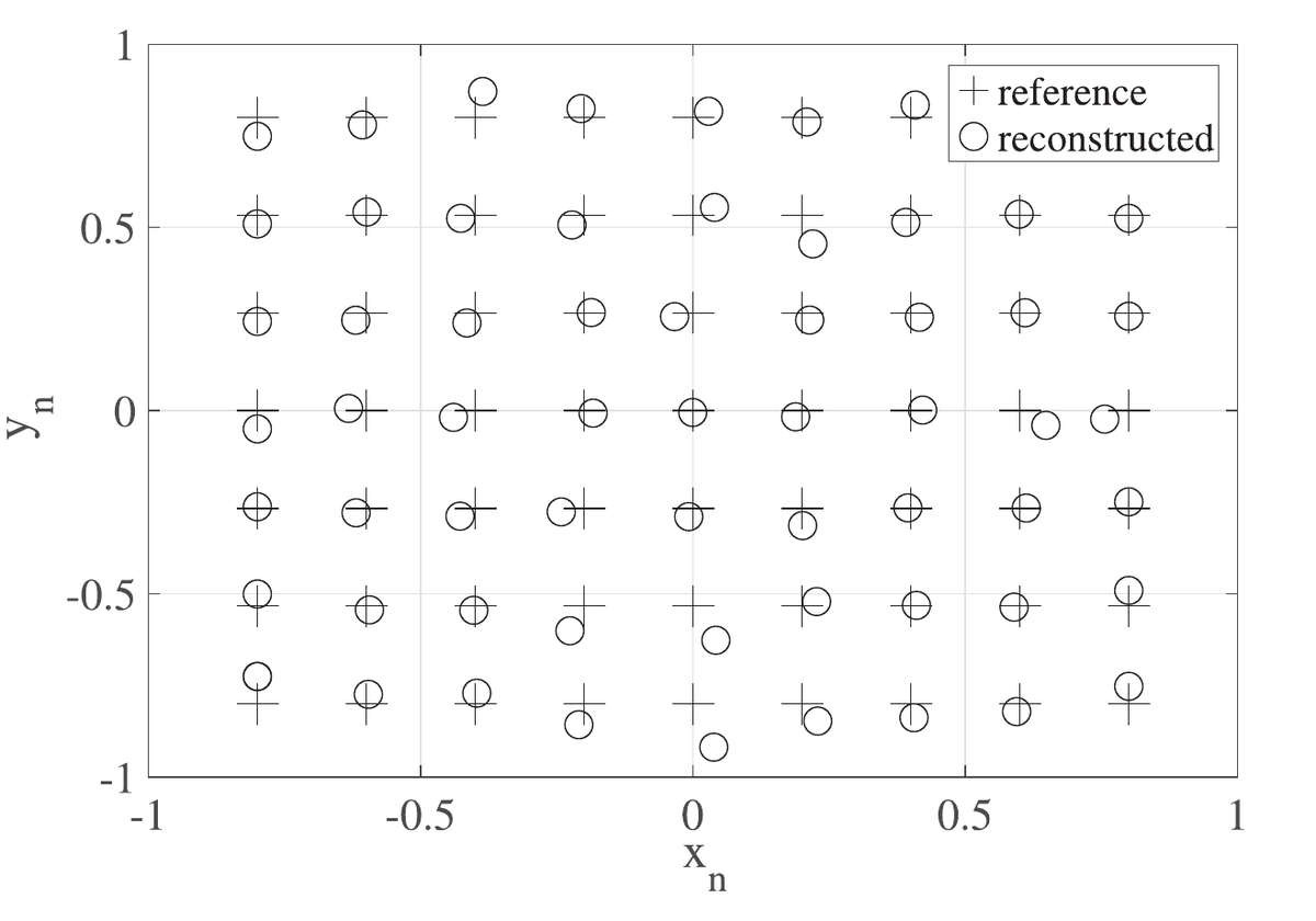
In simulation, the reconstruction error in the central region of the mat was below 0.3 % of the normalised coordinates (see Figure 5). Validation measurements on twelve sensor mats with a 7 × 9 grid of reference positions (see Figure 6) yielded a mean error of 1.23 ± 0.59 mm in the central region, where the PCA muscle is located, and about 1.5 to 1.6 mm near the electrode pins. Since the optimal stimulation area in the PCA muscle measures about 9 × 18 mm, this accuracy is well suited for hotspot training.
Simulator
Virtual component
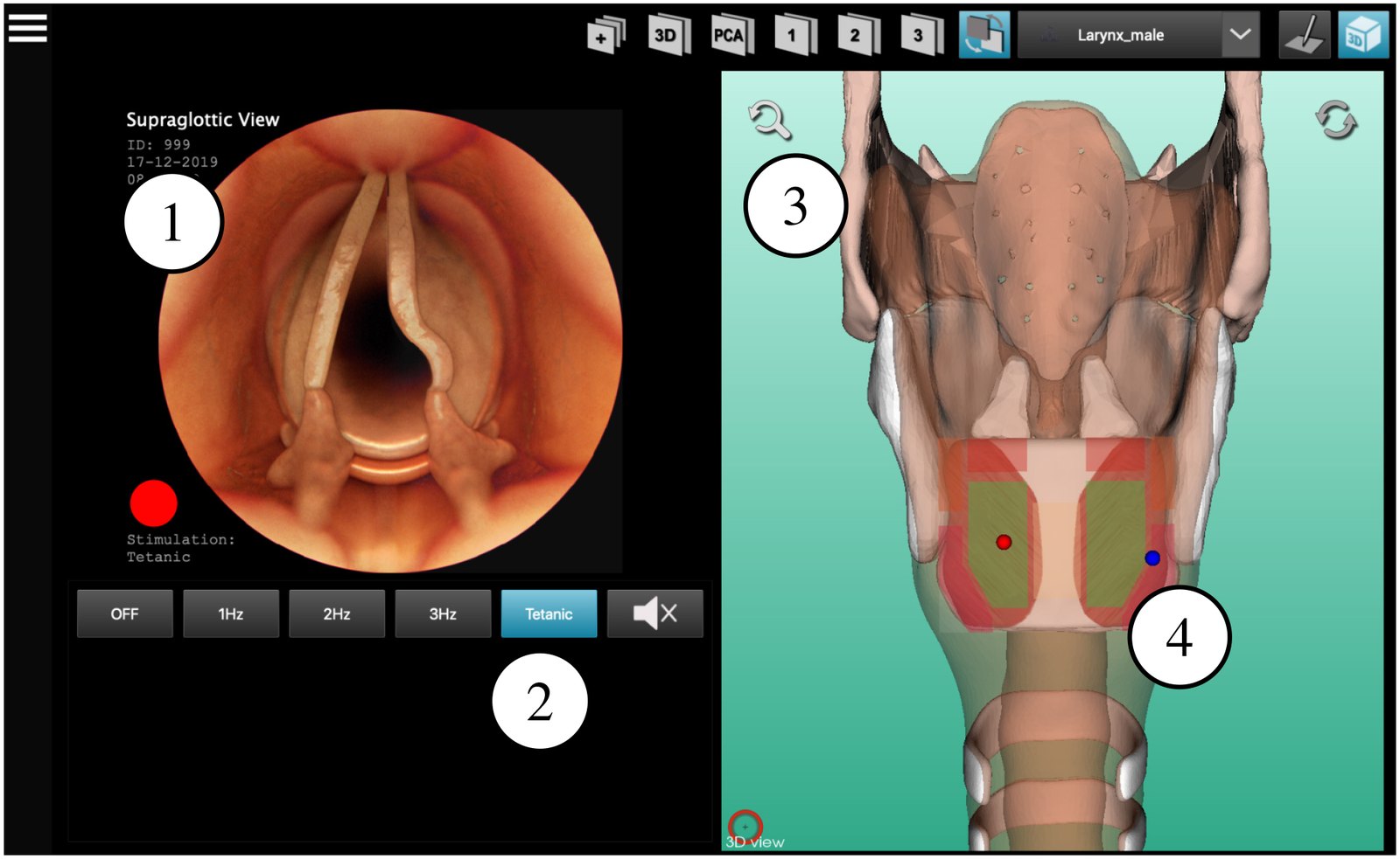
The simulator software (Qt and VTK, operated by touch screen) connects the physical phantom with a virtual scene (see Figure 7). While the LPIT is tunnelled under the synthetic mucosa, a live endoscope view is shown. After penetrating the cricoid, a virtual view of the vocal folds reacts to the electrical stimulation, which is controlled just as in the real intervention: at the hotspot, the vocal folds open, elsewhere they show adverse or mixed reactions. A 3D view of the larynx supports learning the anatomy and shows the positions of both electrode tips in the PCA muscle, with target areas coloured green and sub-optimal areas orange or red.
Physical component
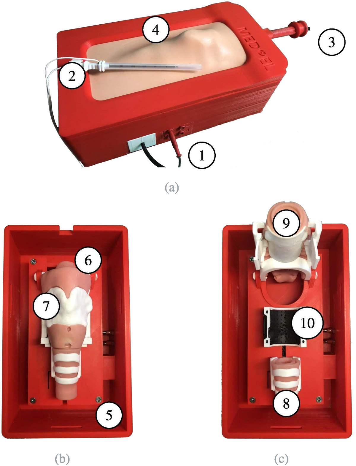
The physical simulator houses the artificial larynx, the sensor electronics and the microcontroller in a compact 3D-printed case (see Figure 8). Two LPITs can be connected; their outer cannula is electrically insulated from the trocar and detects insertions that go too deep, representing a penetration of the oesophagus. A USB endoscope provides sub- and supraglottic views. Structures that are not penetrated are made of durable materials, while the wear parts, the artificial cricoid and the sensor mat, can be replaced in less than two minutes. We recommend replacing them after 20 to 30 simulations.
Final setup
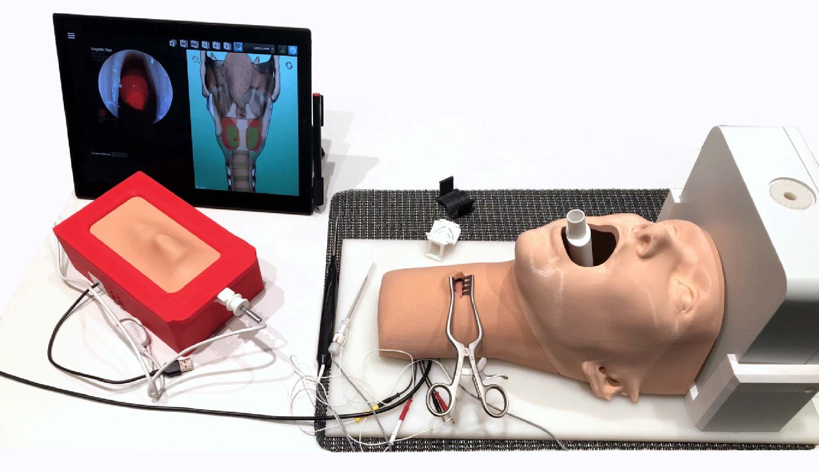
The simulator is available in two versions (see Figure 9): a compact, mobile box version, and a high-fidelity version with a head and neck replica, a laryngeal mask for using a medical endoscope, and additional electromagnetic tracking of the instruments, which records the complete tool path for a detailed debriefing. Both share the same inner hardware for haptics and position detection, and in both, real surgical instruments such as spreaders, forceps and needle holders can be used, and sutures can be placed. The success of a training session can be read directly from the final electrode positions, and its quality from the number of retractions and too deep insertions.
Outlook
Smart Artificial Soft Tissue is a general building block for hybrid simulators: wherever conductive instruments are inserted into tissue, such as in biopsy or needle insertion procedures, it provides continuous, two-dimensional position detection inside haptically realistic artificial anatomy, together with an automatic, objective assessment that many physical simulators lack.
Partners and funding
This project was carried out in cooperation with MED-EL Elektromedizinische Geräte Gesellschaft m.b.H. (Innsbruck), the Division of Soft Matter Physics at Johannes Kepler University Linz, the Institute for Biomechanics of BG Unfallklinik Murnau and Paracelsus Medical University Salzburg, the Department of Otorhinolaryngology at SRH Wald-Klinikum, and Innsbruck University Hospital. It was supported by the Austrian Research Promotion Agency (FFG, project 845436) and by MED-EL.
Figures from [1] licensed under CC BY 4.0.
References
- (2023): Smart Artificial Soft Tissue - Application to a Hybrid Simulator for Training of Laryngeal Pacemaker Implantation. In: IEEE Transactions on Biomedical Engineering, vol. 70, no. 2, pp. 735–746, 2023, ISSN: 0018-9294, (Publisher Copyright: © 1964-2012 IEEE.).
- (2023): Open Source Sensor Interface for Soft Detectors in Surgical Simulators. In: IEEE Access, vol. 11, pp. 49650–49664, 2023, ISSN: 2169-3536, (Publisher Copyright: © 2013 IEEE.).