
MEDUSA
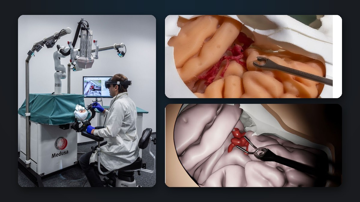
MEDUSA (Medical EDUcation in Surgical Aneurysm clipping) developed a patient-specific mixed-reality simulator for cerebral aneurysm clipping. It combines a physical skull and brain phantom with virtual blood vessels and aneurysms, real surgical instruments and a blood flow simulation that evaluates every clipping strategy immediately [1].
Motivation
Clipping of cerebral aneurysms is one of the most demanding procedures in neurosurgery. Since many aneurysms are now treated endovascularly, open surgery is increasingly reserved for complex, high-risk cases, leaving residents and young neurosurgeons with fewer opportunities to learn it. Realistic simulation can close this gap, both for training and for planning the operation on a specific patient beforehand.
Our contribution
Within the MEDUSA consortium, ReSSL developed the hydraulically actuated brain lobes, integrated them into the hybrid simulator, and contributed to its validation.
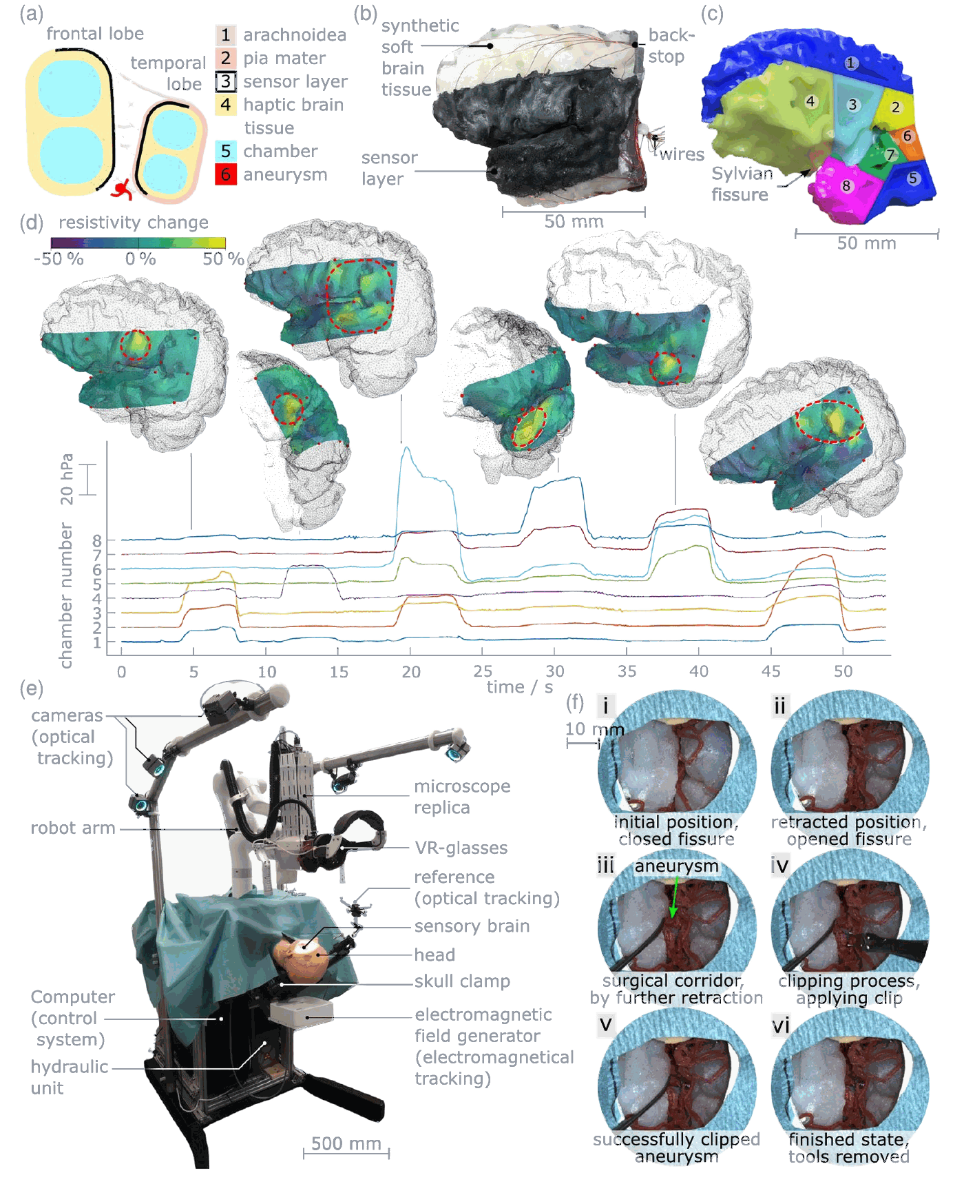
- Dynamic brain phantom: the silicone brain lobes contain fluid-filled chambers that move the frontal and temporal lobe, so the Sylvian fissure can be widened or closed as during surgery. An embedded carbon black silicone sensor layer detects where and how strongly the brain tissue is stressed (see Figure 1 and our project Validation).
The simulator
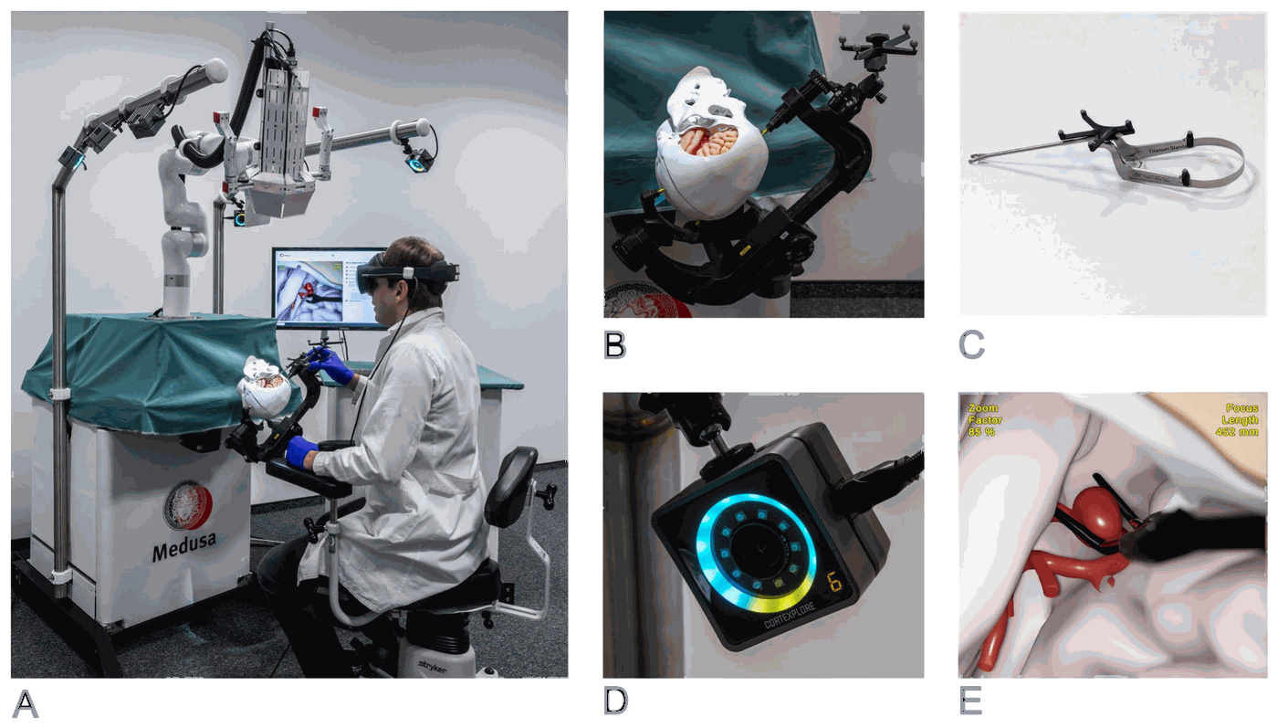
The neurosurgeon sits on a surgical chair and works on a life-sized 3D-printed skull with a pterional craniotomy, mounted in a standard head clamp (see Figure 2). Inside lies a silicone phantom of the left brain hemisphere with the frontal and temporal lobe and the Sylvian fissure in between, so instrument trajectories are restricted just as in reality. A camera on a robotic arm acts as surgical microscope; the trainee sees the operating field through VR glasses, while the same view is shown on a monitor for instructors.
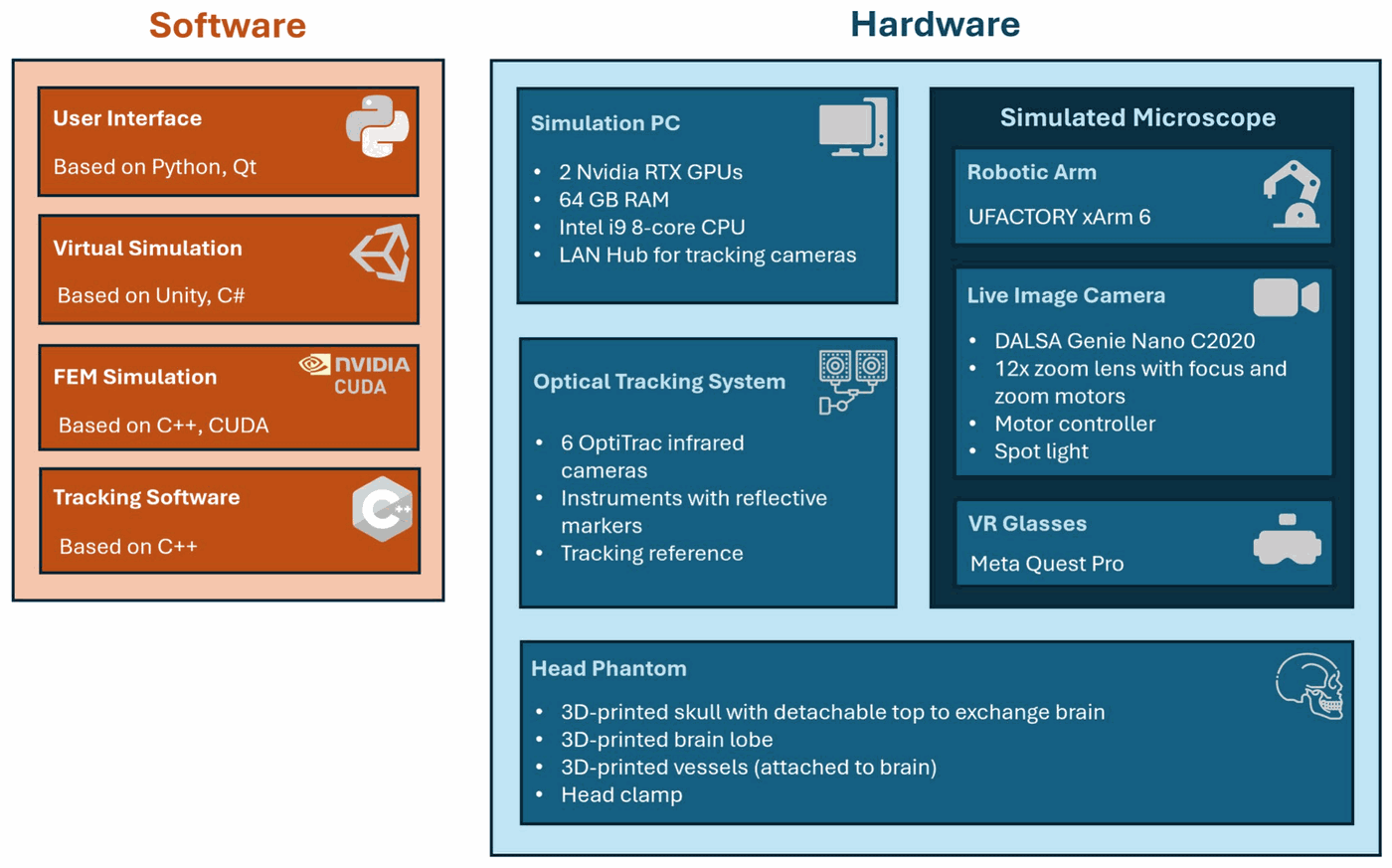
A real clip applier carries reflective markers and is tracked by six infrared cameras with an accuracy of up to 0.1 mm. The clip itself exists only in the virtual world, chosen from a library of more than 50 clip types. When it touches the virtual aneurysm, a finite element model deforms the vessel wall in real time (see Figure 3 for the hardware and software components).
Physical meets virtual
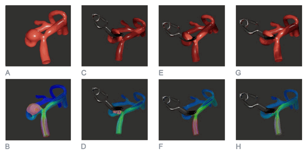
The physical parts provide realistic haptics and restrict the surgical trajectory, while the virtual part allows non-destructive, unlimited repetitions on patient-specific anatomy (see Figure 4). After clipping, a blood flow simulation shows whether the aneurysm is completely occluded or whether a residual aneurysm or a critical stenosis of a parent vessel remains (see Figure 5). Even an intraoperative rupture can be simulated and managed with a temporary clip.
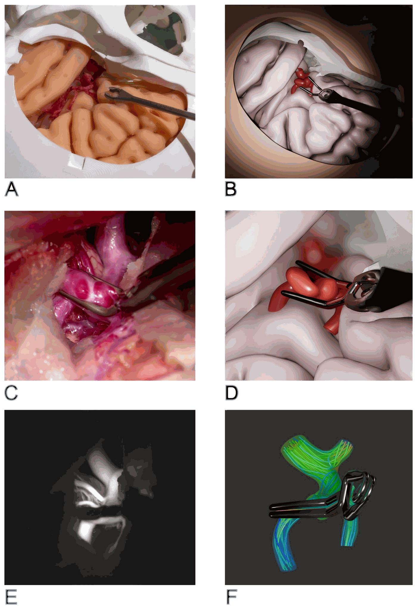
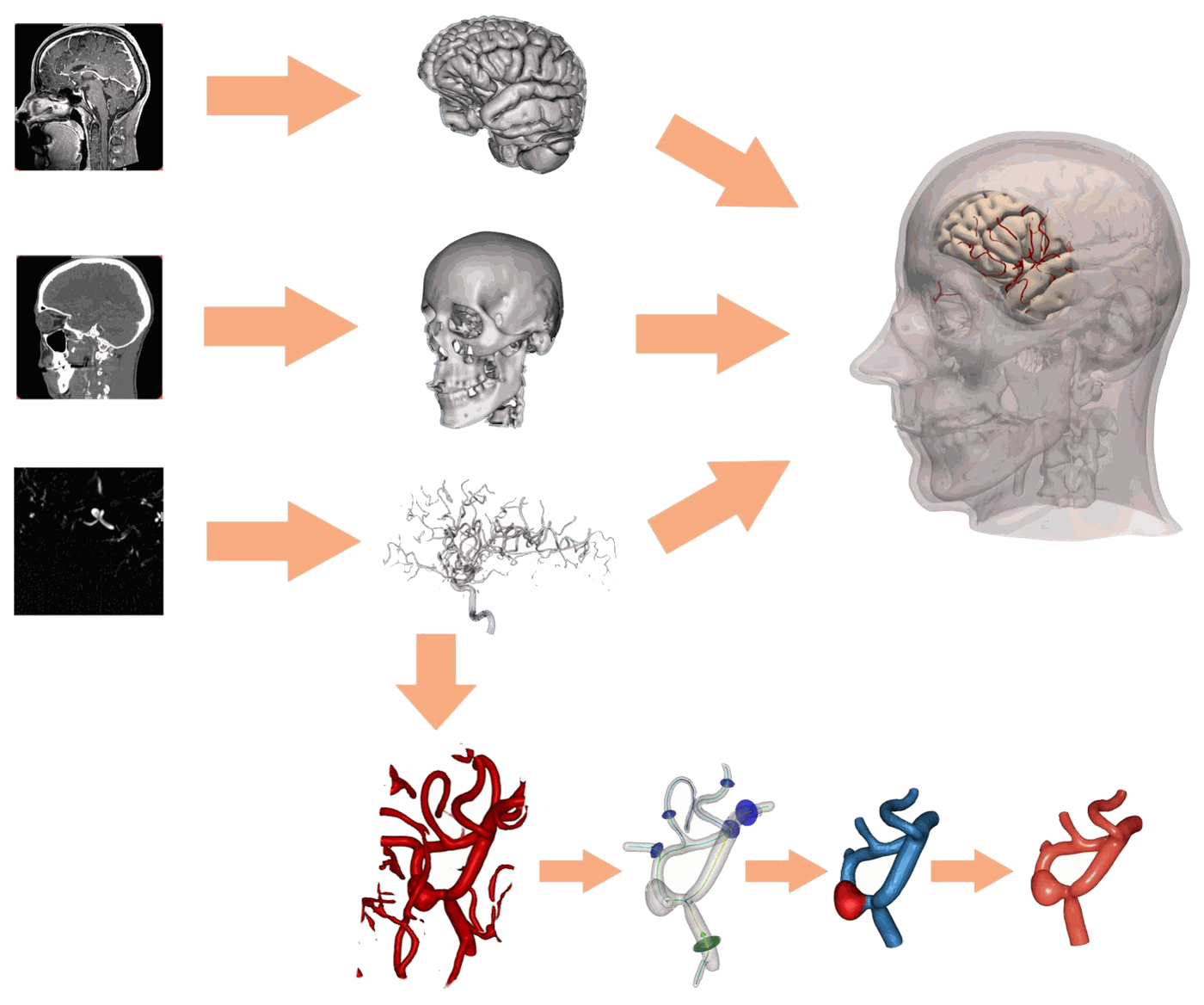
Patient-specific models are created from CT, MRI and angiography data with a pipeline combining AI-based and conventional image processing, which delivers all data needed for a simulation in less than 15 minutes (see Figure 6). This makes it possible to rehearse the operation on “tomorrow’s patient”, even in emergencies.
Validation
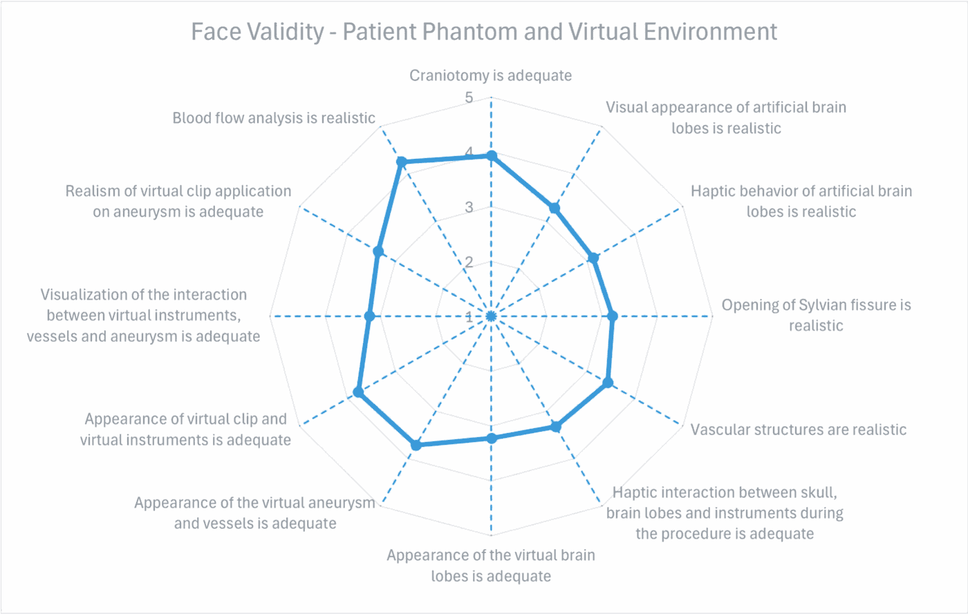
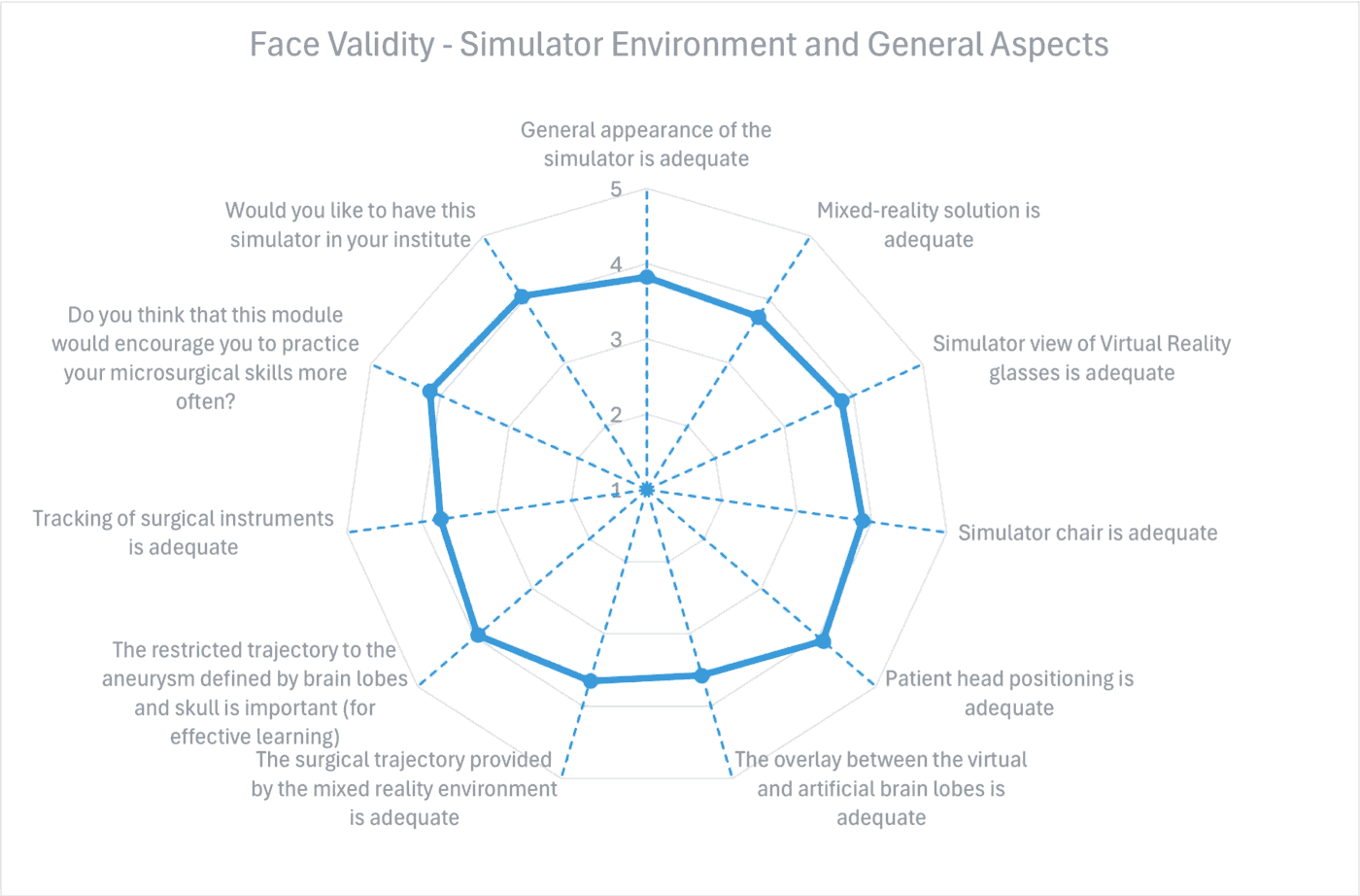
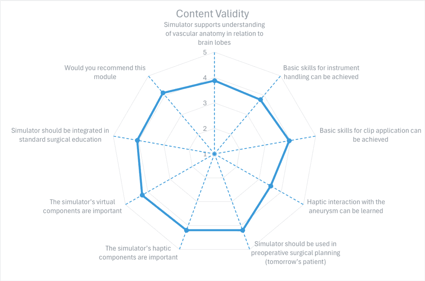
The simulator was externally validated with 40 neurosurgeons, from novices to experts with more than 15 years of experience, at the Zurich Aneurysm Clipping Course (Tübingen 2024, n = 22) and the 11th Annual EANS Vascular Section Meeting (Marseille 2024, n = 18). In a 32-item questionnaire on a 5-point scale, face and content validity were rated with mean scores between 3.13 and 4.25 (see Figures 7–9).
- The blood flow simulation received the highest rating for realism (4.25).
- Participants agreed the simulator should be used for preoperative planning (4.20) and in standard surgical education (4.05).
- Both the physical (4.20) and the virtual components (4.25) were considered important.
- Basic skills for clip application can be achieved on the simulator (3.95).
Outlook
Next steps include a second tracked instrument for bimanual techniques such as suction and dissection, multiple clips per aneurysm, simulated interaction between instruments and the brain lobes, and the validation of the module for splitting the Sylvian fissure, as well as studies on construct and predictive validity.
Project partners
MEDUSA ran from July 2019 to June 2024 as a lead project of the programme “Innovatives OÖ 2020” of the Province of Upper Austria (grant 872604), with a total budget of €3.05 million, coordinated by RISC Software GmbH. Further information is available on the project website.
- alpha medical concepts e.U.
- cortEXplore GmbH
- eulerian-solutions e.U.
- EVO-tech GmbH
- FH OÖ Forschungs & Entwicklungs GmbH (Research Group for Surgical Simulators Linz)
- Johannes Kepler University Linz – Institute of Polymer Product Engineering
- Johannes Kepler University Linz – Institute of Polymer Science
- Kepler University Hospital Linz – Department of Neurosurgery
- LIFEtool gemeinnützige GmbH
- Netural GmbH
- PROFACTOR GmbH
- RISC Software GmbH (coordinator)
- R’n’B Consulting GmbH
Figures 2–9 from [1]; Figure 1 from [2]. Both licensed under CC BY 4.0; backgrounds removed and colours adapted for this website. Partner list: RISC Software.
References
- (2025): Development and external validation of a mixed-reality aneurysm clipping simulator. In: Neurosurgical Review, vol. 48, no. 1, 2025, ISSN: 0344-5607, (Publisher Copyright: © The Author(s) 2025.).
- (2024): Carbon black silicone composite in surgical simulation - A detailed electromechanical characterization. In: Sensors and Actuators, A: Physical, vol. 370, pp. 115262, 2024, ISSN: 0924-4247, (Publisher Copyright: © 2024 The Authors).