
Fetal Heart Phantom
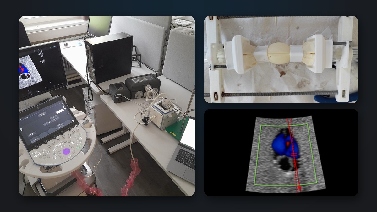
FHSimApUs (Fetal Heart Simulator for the Development and Optimization of Ultrasound Imaging Algorithms) developed an active ultrasound phantom of a beating fetal heart. Together with GE Healthcare, the phantom was used to develop and validate new imaging algorithms for fetal echocardiography.
Motivation
Imaging the fetal heart is one of the most demanding tasks in prenatal ultrasound: the heart is tiny, beats at up to about 180 beats per minute and lies deep inside the mother’s body. Developing and testing ultrasound algorithms for it requires reproducible conditions, which recordings from real pregnancies cannot provide. A phantom that beats at an adjustable rate, pumps fluid through its chambers and looks like real tissue in ultrasound makes such algorithms testable and comparable, and it can also serve for quality assurance and training.
Tissue-mimicking materials
The phantom requires materials that behave acoustically like human tissue. We built a measurement setup, later extended by a hydrophone, that determines the speed of sound, the attenuation and the acoustic nonlinearity parameter B/A of tissue-mimicking materials; the method for estimating B/A was published in the Journal of the Acoustical Society of America [1].
A rigid agar phantom restricted the movement of the heart too much, so we developed a soft, emulsion-based material (water, soy lecithin, corn oil, glucose syrup, xanthan, silicon carbide and a preservative) that lets the heart move freely, keeps much longer than milk-based recipes, and matches the target values for the surrounding tissue: a speed of sound of 1540 m/s and an attenuation of 0.5 dB/MHz/cm.
The beating fetal heart
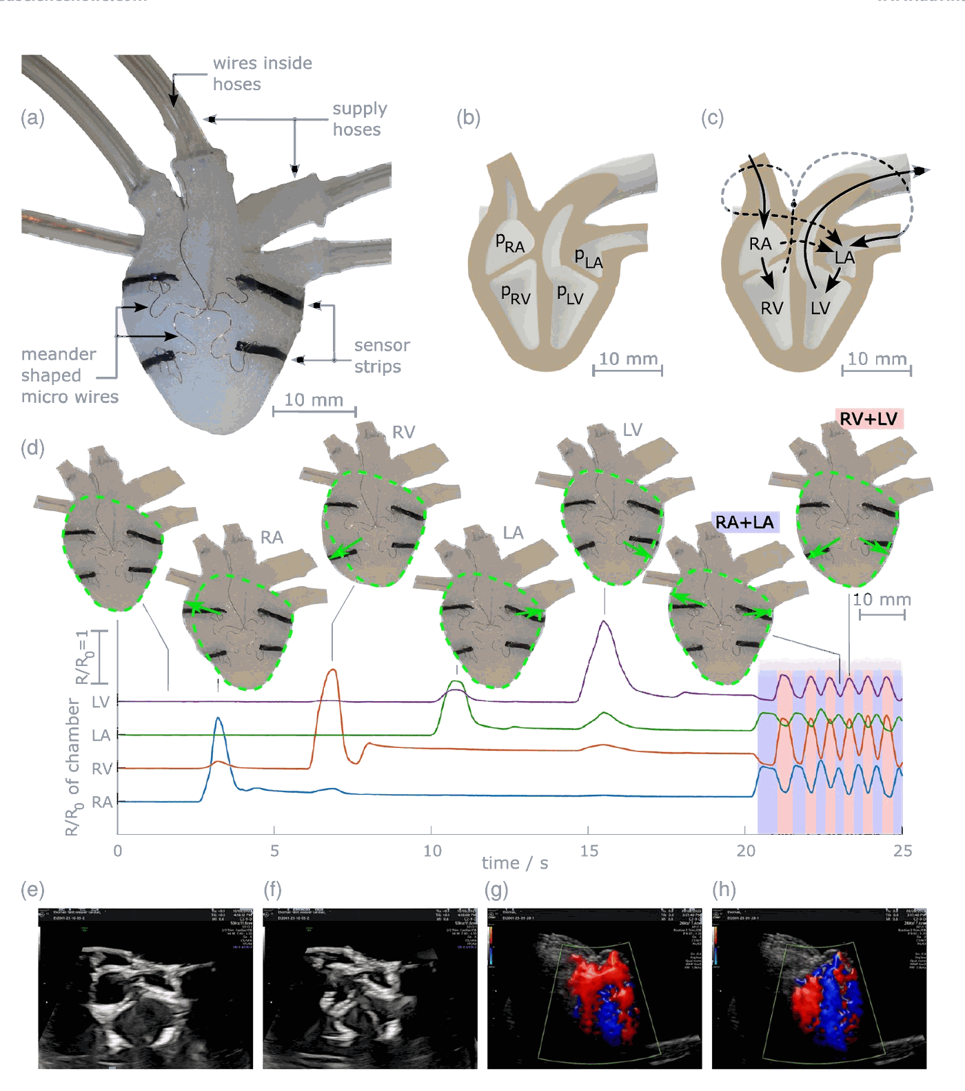
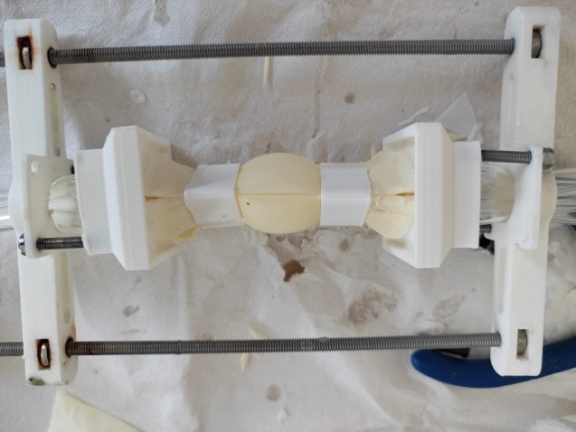
The artificial heart has four chambers, corresponding to the right and left atrium and ventricle. A first, cast version was tested with soft sensor strips as part of our Dynamic Tactile Synthetic Tissue (see Figure 1) [2]. Its walls, however, attenuated the ultrasound too strongly. The final heart is therefore built from thin latex layers mounted on a 3D-printed frame, with clips that create realistic swirling of the flow inside the chambers (see Figure 2), which significantly improved the ultrasound image.
Phantom and drive system
The heart is embedded in the tissue-mimicking material inside an acrylic housing (see Figure 3). Its scanning windows are sealed with natural rubber film instead of the originally planned PET film: latex differs only slightly in acoustic impedance from the surrounding material, which minimises reflections, and its elasticity is ideal when the probe is pressed on automatically, for example by a robot. The supply hoses leave the housing sideways so they don’t interfere with the four-chamber view, and a flushing program removes air bubbles from the system.
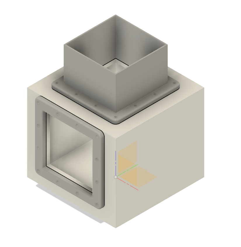
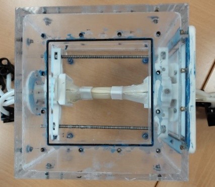
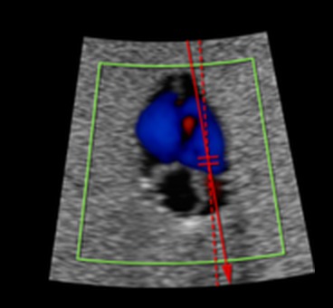
Figure 3: Phantom housing as CAD drawing (left), prototype with the latex heart before filling with tissue-mimicking material (middle), and colour Doppler image of the beating heart, showing the flow through the chambers (right).
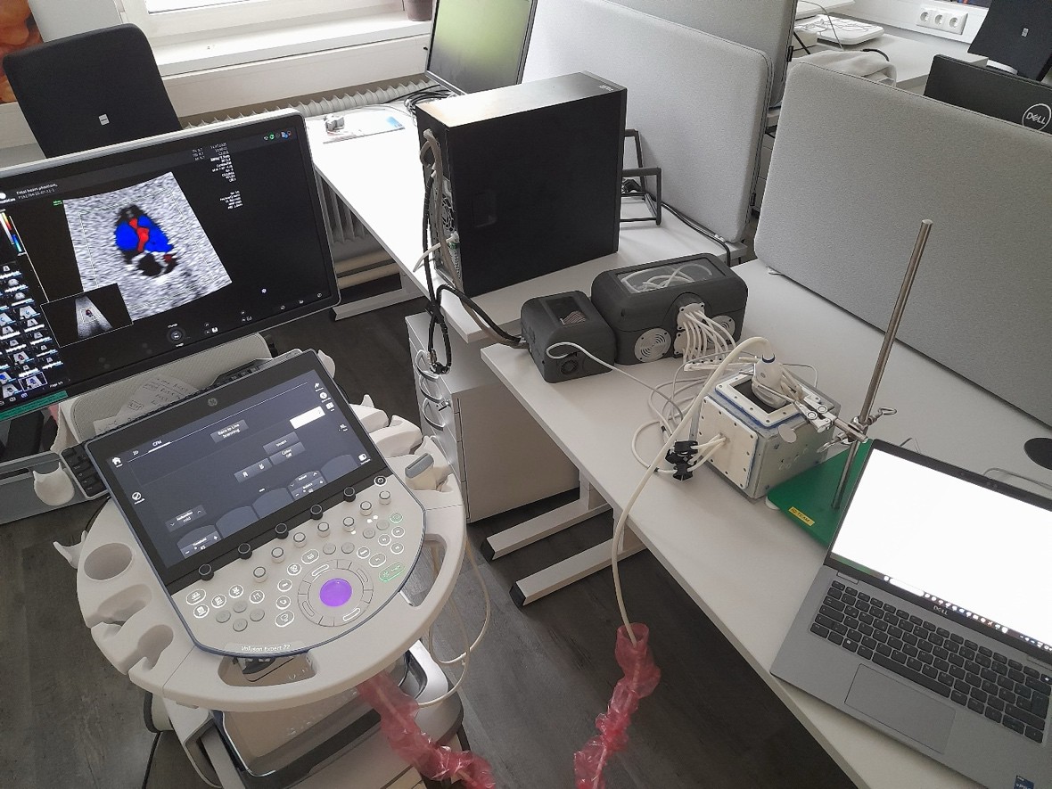
A hydraulic system with pressure and vacuum tanks, valves and pressure sensors drives the heart chambers. It is controlled by a Raspberry Pi, and the user sets the heart rate, the chamber pressures and the valve timing in a MATLAB app (see Figure 4). All connections can be unplugged, and wear parts such as the rubber windows or the heart itself can be exchanged easily.
Validation and imaging algorithms
The phantom was developed iteratively until it met all functional and sonographic requirements, and then handed over to GE Healthcare. There, the heart rate set in the control software matched the one measured in the ultrasound system exactly, for example 174 beats per minute in M-mode and 119 beats per minute in colour Doppler, which also showed the correct direction of flow through the chambers.
Using the phantom as a reproducible test object, GE Healthcare developed new acquisition and processing methods for spatio-temporal image correlation (STIC/eSTIC) of the fetal heart, based on retrospective transmit beamforming, as well as a nonlinear temporal filter that reduces noise and motion artefacts. Validated on the phantom and on real fetal data, they improved spatial and temporal resolution at acquisition frame rates above 200 Hz, without affecting the accuracy of heart rate measurement.
Results
- A beating, four-chamber fetal heart phantom with adjustable heart rate and pressures, validated in B-mode, M-mode and colour Doppler, now used by GE Healthcare for testing, validation and benchmarking.
- A measurement setup and tissue-mimicking materials with characterised speed of sound, attenuation and nonlinearity.
- Two journal publications [2], [1] and four master’s theses.
Outlook
Building on this project, Thomas Thurner is founding a start-up for surgical simulators, with ultrasound phantoms for image quality assurance and medical training as one possible field. Together with University of Applied Sciences Upper Austria and RISC Software GmbH, the follow-up project HyTra (FFG, Expedition Zukunft – START) develops the technical basis for a simulator for surgery on the unborn child.
Partners and funding
FHSimApUs was carried out by the Research Group for Surgical Simulators Linz (Andreas Schrempf, Lisa Stefely, Thomas Thurner) together with GE Healthcare, Zipf, from October 2022 to October 2025. It was funded by the Province of Upper Austria within the programme “Digital Health – The Digital Patient Journey” (FFG project number 41634460).
Figure 1 taken from [2], licensed under CC BY 4.0.
References
- (2026): A model-based approach for B/A estimation using the finite amplitude insertion-substitution method. In: Journal of the Acoustical Society of America, vol. 159, no. 2, pp. 1416–1429, 2026, ISSN: 0001-4966, (Publisher Copyright: © 2026 Author(s).).
- (2024): Dynamic Tactile Synthetic Tissue: from Soft Robotics to Hybrid Surgical Simulators. In: Advanced Intelligent Systems, vol. 6, no. 12, 2024, ISSN: 2640-4567, (Publisher Copyright: © 2024 The Author(s). Advanced Intelligent Systems published by Wiley-VCH GmbH.).