
CranioSim
This project aims to develop a surgical simulator for training cranial bone graft harvesting techniques.
Motivation
The first transplantation of a bone graft was reported in 1668 by the Dutch surgeon Job van Meekeren, who transplanted a graft harvested from a dog’s skull into a soldier’s cranial bone [1]. Today, bone grafts are harvested from various donor sites, such as the iliac crest, tibia, maxilla, mandible, ribs, sternum and calvarial bone [2]. The calvaria has become a popular donor site for autologous grafts, especially in cranio-maxillofacial surgery [3].
In 1982, Tessier first reported the use of autogenous calvarial bone as graft material for facial reconstruction [4]. Cranial bone offers numerous advantages as a graft material: reduced postoperative pain at the donor site, a scar well hidden beneath the scalp, a shorter hospital stay and, in maxillofacial surgery, a single surgical field. In addition, calvarial grafts undergo less resorption and may revascularise more rapidly [5].
Because the skull is convex rather than flat, harvesting a suitable graft from the outer table is challenging. During harvesting, penetration of the cranial vault must be avoided at all costs [3]. Disadvantages of this donor site include the risk of a dural tear, contour deformities of the skull and the small amount of cancellous bone available for harvest [2].
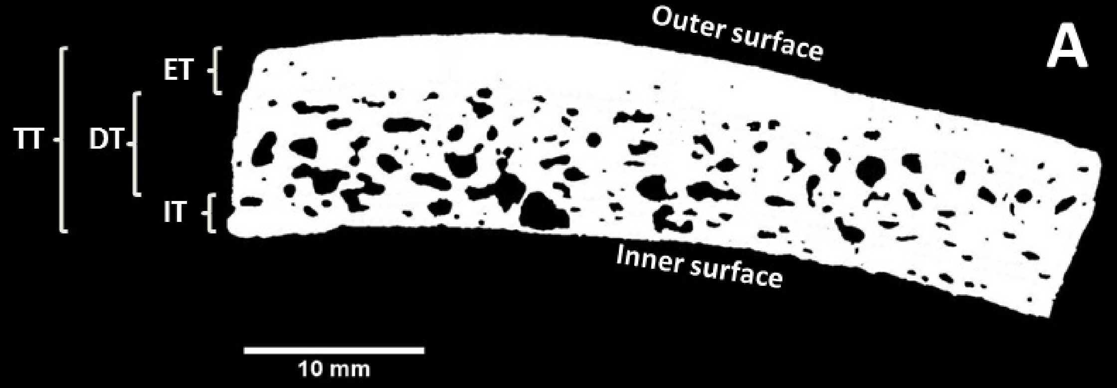
The bone of the skull vault consists of three layers: the outer and inner tables (tabula externa and interna) consist of compact bone, whereas the middle layer, the diploë, consists of spongy, trabecular bone. The parietal bone is the preferred harvest site for calvarial grafts because of its thickness [5].
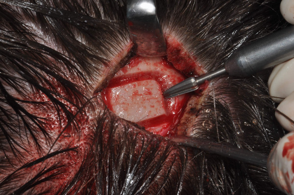
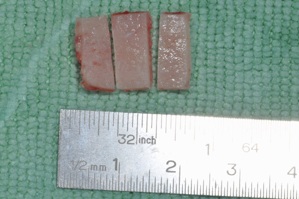
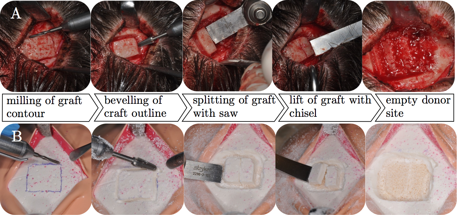
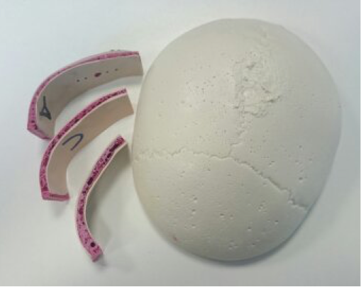
A typical technique for harvesting calvarial bone (see Figure 3) is performed as follows: after the scalp has been incised and held open with clamps, the outline of the graft is cut with a cutting burr, to a depth that depends on the thickness of the calvarial bone. An oscillating saw is then advanced into the diploic space. Finally, a curved osteotome cuts the remaining diploic connections between the outer and inner table [3], releasing the autologous graft (see Figure 2).
Simulator
Artificial cranial bone
To build a simulator for training parietal graft harvesting, polyurethane (PU)-based bone surrogate composites enriched with mineral fillers were developed (see Figure 4) [6], [7], [8]. To mimic spongy bone covered by a cortical layer, as in the human skull, four material compositions were created. For an open-cell cancellous structure, the PU resin was supplemented with water as a blowing agent and a cell stabiliser. This cancellous PU mixture was further enriched with one of two fillers, calcium phosphate (CaP, Ca3(PO4)2) or calcium carbonate (CaC, CaCO3), in amounts of 50 % (CaP050, CaC050) and 100 % (CaP100, CaC100) of the total mass to vary structure and hardness. After curing, the cancellous blocks were cut flat with a band saw and coated with a second, cortical mixture of PU resin and mineral fillers, spread evenly with a spatula to a layer thickness of 1.5 mm. This corresponds to the thickness of the human outer table of about 1.6 ± 0.2 mm [9].
Patient phantom
A patient phantom for training cranial bone graft harvesting was built from these bone surrogates. The artificial skull cap was placed on a silicone base with a brain-shaped bulge to prevent slipping and covered with layers of artificial soft tissue (muscle and skin). After skin incision and retraction of the soft tissues, surgeons can train the same procedural steps as in a real parietal graft harvest. The simulator prototypes were tested by two experienced surgeons (see Figure 5).
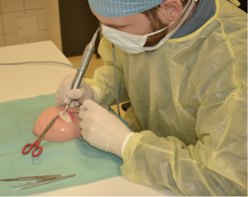
The following video shows a surgeon harvesting a cranial graft on the simulator.
Validation
The simulator, i.e. the customised patient phantom, was validated in two ways: by comparing characteristic forces during drilling, milling and sawing with those measured in human parietal bone, and by an evaluation study assessing face, content and construct validity.
Surgical machining
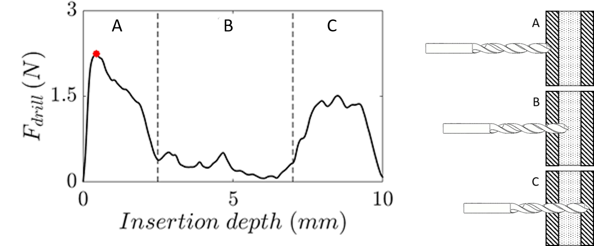
Following the surgical procedure of a split-thickness tabula externa graft harvest, the maximum puncture forces of drill and mill heads penetrating the tabula externa were used to compare the machining characteristics of artificial and human skull bone (see Figure 6). In addition, the maximum force during cancellous sawing, reached when the saw blade is fully inserted into the cancellous bone, was identified as a characteristic parameter.
Drilling
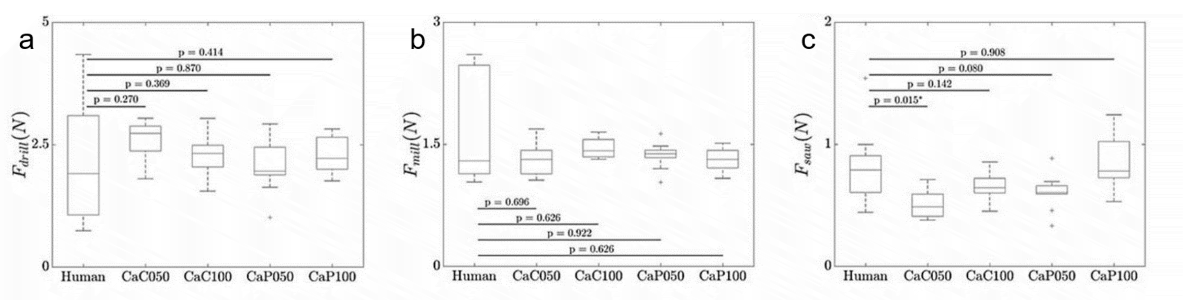
Among the bone surrogates, the highest axial drilling forces were recorded for CaC050 (2.5 ± 0.2 N), compared with 1.8 ± 0.5 N for the human specimens. On average, the maximum drilling forces of the artificial bone materials were 18 % (CaP050), 20 % (CaP100), 42 % (CaC050) and 32 % (CaC100) higher than those of the human bones. The Shapiro-Wilk test showed normal distributions for all drilling measurements, and variances were homogeneous across all groups. No significant differences were found between the human specimens and the bone surrogates (see Figure 7).
Milling
The lowest axial milling forces were found for CaC050 (1.3 ± 0.1 N), clearly below those of human parietal bone (1.7 ± 0.3 N). Overall, the mean maximum milling forces were 18 % (CaP050), 21 % (CaP100), 25 % (CaC050) and 16 % (CaC100) lower than those of the human specimens. As the Shapiro-Wilk test indicated a non-normal distribution for the human specimens, a Mann-Whitney U test was used to compare human and artificial specimens; all bone surrogates showed high p-values (see Figure 7).
Sawing
The Shapiro-Wilk test indicated normal distributions for all sawing measurements, and Levene’s test showed homogeneous variances. Therefore, an unpaired Student’s t-test was used to compare human and artificial bone. The lowest sawing forces were found for CaC050 (0.5 ± 0.1 N). The mean maximum sawing forces of CaP050, CaP100, CaC050 and CaC100 were 32 %, 11 %, 43 % and 26 % lower than those of the human bone samples (see Figure 7).
Anatomical layers
To create an artificial skull cap with anatomically realistic bone layers, the thicknesses of the tabula externa, diploë and tabula interna, as well as the total thickness of parietal bone samples, were measured (see Figure 8). The results served as a reference for developing the bone layers of the artificial skull cap (ASC), in particular for estimating the amount of material needed to mould each layer. All human specimens were scanned with a micro-computed tomography scanner (µCT 80, Scanco Medical, Brüttisellen, Switzerland) at 70 kV, 114 µA and an integration time of 200 ms, resulting in a slice thickness of 0.09 mm.
For all samples, the thicknesses of the inner and outer cortical layers and the total thickness were measured. The thickness of the diploic space was then calculated by subtracting the cortical layer thicknesses from the total bone thickness.
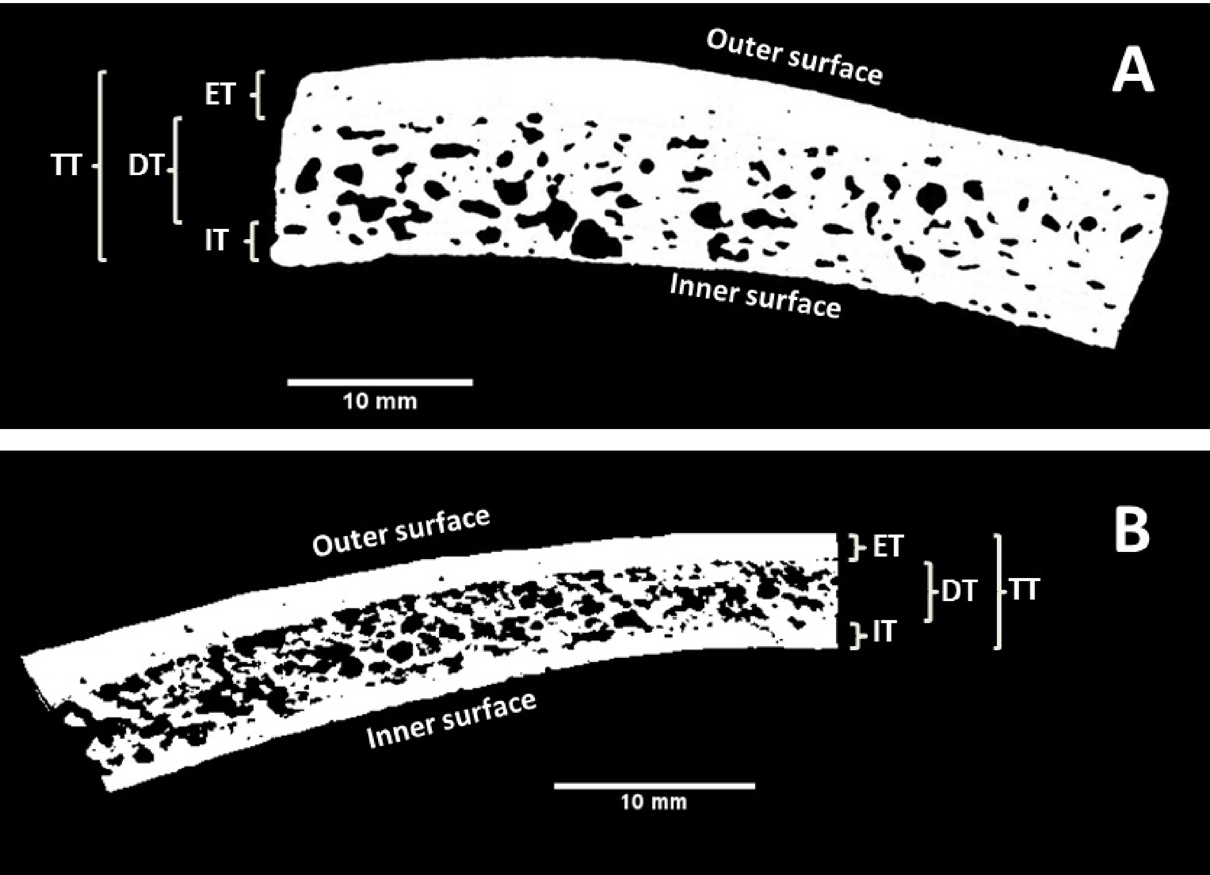
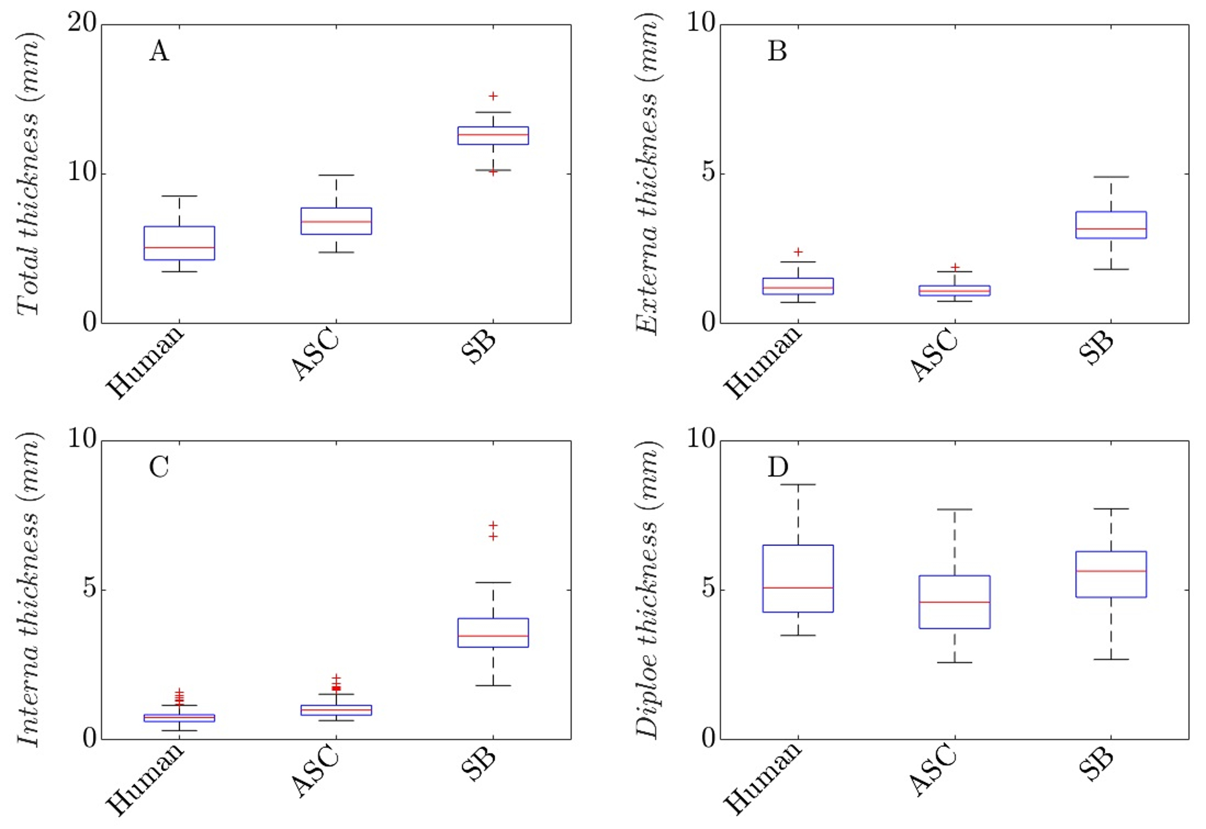
The results are summarised in Figure 9. The human bone samples had a total thickness of 5.31 ± 1.24 mm. The total thickness of the artificial skull cap (ASC) was 6.94 ± 1.20 mm, i.e. 30.1 % larger. The Sawbones (SB) skull samples had a total thickness of 12.51 ± 1.14 mm and were thus more than twice as thick (+135.4 %) as the human samples. The tabula externa of the human bones measured 1.26 ± 0.34 mm. The artificial skull caps were similar (1.15 ± 0.25 mm, only about 9 % thinner), whereas the SB samples were more than 165 % thicker (3.35 ± 0.73 mm).
Similar results were found for the tabula interna. The average interna thickness of human parietal bone was 0.76 ± 0.22 mm, whereas ASC (1.07 ± 0.34 mm, +40.7 %) and SB (3.69 ± 1.18 mm, +385.5 %) were clearly thicker.
Validation study
This study aimed to verify the face, content and construct validity of the novel model-based simulator [10]. Participants were asked to perform a tabula externa graft harvest during a training session on the simulator, and their task performance was analysed with a standardised assessment tool.
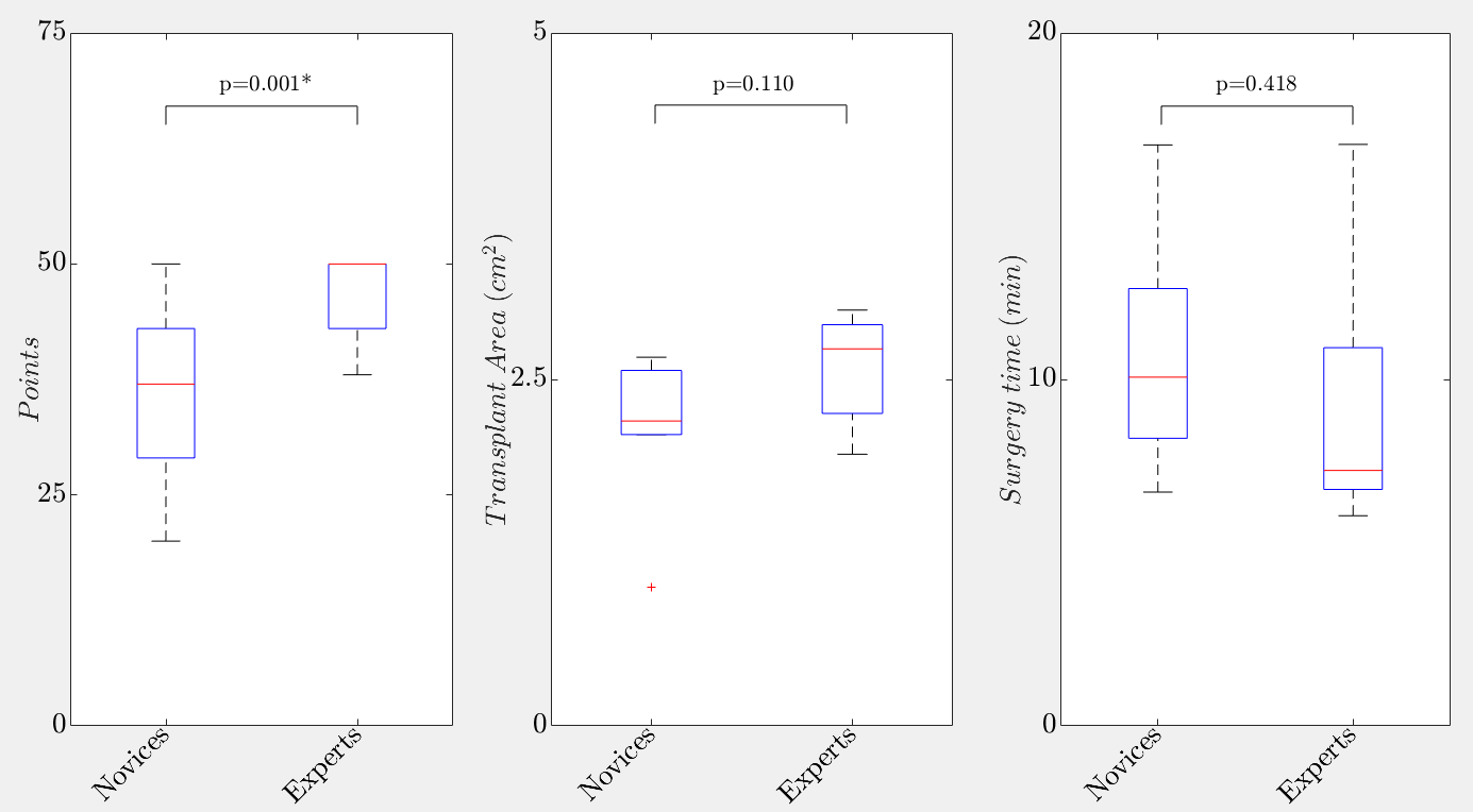
Before the training task, the participants (n = 15) completed questionnaires on demographics and medical experience. Experts and novices then harvested a 2 × 1.5 cm graft, split into three equal parts, from the parietal tabula externa of the simulator.
Participants rated the realism of the simulator (TabSIM) and its suitability as a teaching tool on a 5-point Likert scale. Face validity (realism) was confirmed with an average score of 4.2 out of 5, and content validity (suitability for teaching) with an average score of 4.8 out of 5. In the task performance assessment, the expert surgeons scored significantly better than the novices, confirming the construct validity of the simulator (see Figure 10).
References
- (1988): The history of Bone Grafts. In: Clinical Orthopaedics and Related Research, vol. 226, pp. 292-298, 1988.
- (2010): Bone Graft Harvesting From Distant Sites: Concepts and Techniques. In: Oral and Maxillofacial Surgery Clinics of North America, vol. 22, no. 3, pp. 301-316, 2010, ISSN: 1042-3699.
- (1990): An instrument for harvesting the outer table of the skull. In: Journal of Cranio-Maxillofacial Surgery, vol. 18, no. 4, pp. 179-181, 1990, ISSN: 1010-5182.
- (1982): Autogenous bone grafts taken from the calvarium for facial and cranial applications. In: Clin Plast Surg, vol. 9, pp. 531-538, 1982.
- (1994): Cranial Bone Harvest, Grafting. In: AORN Journal, vol. 59, no. 1, pp. 242-251, 1994, ISSN: 0001-2092.
- (2017): Novel synthetic vertebrae provide realistic haptics for pedicle screw placement. In: Proceedings of the 39th Annual International Conference of the IEEE Engineering in Medicine and Biology Society, 2017.
- (2016): Development of parietal bone surrogates for parietal graft lift training. In: Current Directions in Biomedical Engineering, 2016.
- (2017): Novel Simulator for Cranial Graft Lift Training. In: Proceedings of the IAUP Triennial Conference, 2017.
- (2003): Regional thickness of parietal bone in Korean adults.. In: International journal of oral and maxillofacial surgery, vol. 32, iss. 6, pp. 638–641, 2003, ISSN: 0901-5027.
- (2018): Validation of a Simulator for Cranial Graft Lift Training: Face, Content, and Construct Validity. In: Journal of Cranio-Maxillo-Facial Surgery, vol. 46, no. 8, pp. 1390–1394, 2018.